TL;DR: A 2026 Imaging Neuroscience functional MRI (fMRI) study found that emotional pictures were recalled better than neutral pictures, but amygdala and insula activity appeared tied mainly to arousal rather than whether memories were negative or positive.
Key Findings
- Large fMRI sample: The main imaging analysis used 1,006 healthy young adults from a single center.
- Memory behavior: Participants recalled more positive and negative pictures than neutral pictures.
- Arousal result: Amygdala and insula effects were no longer significant after controlling for subjective arousal.
- Valence pattern: Negative and positive memory enhancement shared occipitotemporal regions but also had distinct networks.
- Caution: The study used picture encoding and free recall in healthy young adults, not clinical trauma or autobiographical memory.
Source: Imaging Neuroscience (2026) | Amini et al.
Emotional events are often remembered better than neutral ones.
That effect can come from at least two ingredients: arousal, meaning how activating or intense something is rated, and valence, meaning whether it is negative or positive.
Amini and other researchers used a large functional MRI, or fMRI, sample to separate those ingredients.
Participants saw negative, neutral, and positive pictures during scanning, then completed an unannounced free-recall test after leaving the scanner.
The study supports a more specific memory model. Amygdala and insula activity appeared important for emotional arousal, while negative and positive memory enhancement also recruited partly distinct cortical networks.
That distinction is useful because people often talk about “emotional memory” as if it were one thing.
A frightening image, a joyful image, and a merely intense image can all be emotional, but the brain may not treat them as interchangeable.
1,006 Young Adults Completed Emotional Memory fMRI
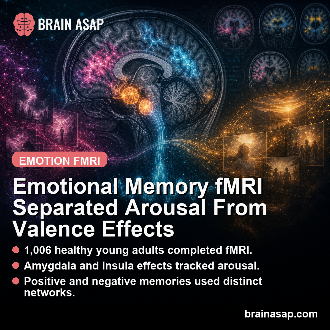
The main neuroimaging analysis used 1,006 healthy young adults. Behavioral analyses included a larger full sample of 1,591 participants.
During scanning, participants viewed 72 pictures across negative, neutral, and positive categories. After scanning, they completed a free-recall task. The researchers then asked which brain activity during encoding predicted later memory.
This is called a subsequent-memory analysis. It compares brain activity for items later remembered versus not remembered, then tests whether that difference is stronger for emotional than neutral items.
The design has two strengths.
First, the sample was unusually large for neuroimaging.
Second, the researchers collected subjective ratings of valence and arousal, which allowed them to test whether a brain region was responding to emotional category or to perceived intensity.
The task was still controlled and simple.
Participants were remembering pictures shown in a scanner, not personal life events.
The design is cleaner for experiment purposes, but it also limits how far the result should be stretched.
Positive and Negative Pictures Were Recalled More Than Neutral Pictures
Participants recalled an average of 30.8 out of 72 pictures. Positive pictures were recalled most often, negative pictures next, and neutral pictures least often.
The recall counts were clear:
- Positive pictures: 12.2 recalled on average.
- Negative pictures: 11.4 recalled on average.
- Neutral pictures: 7.2 recalled on average.
Subjective ratings confirmed the picture categories.
Participants rated negative pictures as negative, positive pictures as positive, and both emotional categories as more arousing than neutral pictures.
Negative pictures were rated more arousing than positive pictures.
The behavioral result is the foundation for the imaging analysis. If emotional pictures had not been remembered better, there would be less reason to ask which encoding signals explained the memory advantage.
The recall pattern also shows why arousal needed to be handled carefully.
Negative pictures were both emotional and more arousing, so a simple negative-versus-neutral comparison could mistakenly label an arousal effect as a negative-valence effect.
Positive pictures also mattered for that reason.
If only negative pictures had been tested, the study could not have separated “emotional” from “negative.” Including both positive and negative images made it possible to ask which findings were shared across emotion and which depended on valence.
Amygdala and Insula Effects Were Arousal-Related
The first emotional-versus-neutral analysis identified regions often seen in emotional memory studies: occipitotemporal visual cortex, anterior cingulate cortex, insula, and amygdala. The study also found an extensive temporoparietal network.
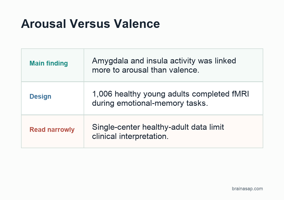
The crucial test came next. After controlling for participants’ subjective arousal ratings, clusters in the amygdala and insula were no longer significant.
That does not make the amygdala unimportant.
It suggests a more specific role: in this task, amygdala and insula activity tracked the arousal part of emotional memory more than the positive-versus-negative valence part.
That finding fits a broader view of the amygdala as a detector of motivational relevance and intensity, not only a “fear center.” It can respond when information is salient, biologically important, or worth prioritizing for memory.
The insula result also fits the arousal interpretation.
The insula is often involved in bodily feeling states and internal awareness, which fits a role in tracking how activating an image was rated by the participant.
Negative and Positive Emotional Memories Used Partly Different Networks
After accounting for arousal, negative and positive memory enhancement still had overlapping and distinct patterns. Shared effects were strongest in occipitotemporal regions involved in visual processing.
The valence-specific pattern was different:
- Negative memory enhancement involved sensory-attentional regions, including lateral occipital and supramarginal areas.
- Positive memory enhancement involved anterior cingulate, superior frontal, parietal, and precuneus regions.
- Neutral memory encoding relied more on scene-related and frontoparietal control regions.
The result argues against treating emotional memory as one undifferentiated system. Arousal, negative valence, positive valence, visual attention, and memory control can each contribute.
The study also helps explain why emotional-memory findings can look inconsistent across papers.
One experiment may use highly arousing negative images, another may use pleasant images, and another may rely on recognition rather than free recall.
Those choices can change which brain systems appear most important.
For quick reading, the split is:
- Arousal: Arousal was most relevant for amygdala and insula effects.
- Valence: Negative and positive valence still showed partially different cortical patterns.
- Neutral memory: Neutral memory relied more on scene and control-related regions.
The Study Refines Emotional Memory Models
The study is valuable because it is large and separates valence from arousal. Many smaller imaging studies cannot cleanly test that distinction, especially when negative pictures are more arousing than positive pictures.
The limits are equally important. The participants were healthy young adults. The memories were laboratory picture memories, not autobiographical events, trauma memories, or clinical symptoms.
Free recall also differs from recognition memory, which is common in other emotional-memory studies. A person may fail to freely recall an image but still recognize it later when prompted.
The practical interpretation: when emotional memories become stronger, the amygdala and insula may be responding to intensity, while positive and negative memory effects can still rely on partly different cortical networks.
That helps explain why “emotional memory” is too broad unless the study specifies arousal, valence, and memory test type.
In mental health contexts, this should not be treated as a direct trauma study. It is better read as a basic neuroscience paper that sharpens the vocabulary.
Before claiming that a brain region supports emotional memory, researchers need to ask which ingredient they mean: intensity, negativity, positivity, attention, or retrieval style.
The study also gives a good example of why subjective ratings belong in emotion research. Brain activity can look valence-specific until researchers account for how intense the stimulus was to the participant.
Without that step, arousal can quietly drive a result that gets described as negative or positive emotion.
Citation: DOI: 10.1162/IMAG.a.1213. Amini et al. Neural correlates of emotional memory enhancement: The role of valence and arousal. Imaging Neuroscience. 2026;4.
Study Design: Functional MRI study separating emotional-memory arousal and valence effects.
Sample/Model: 1,006 healthy young adults completed picture encoding and free recall during fMRI.
Key Statistic: Amygdala and insula effects were mainly arousal-related after subjective arousal was controlled.
Caveat: Picture-recall findings in healthy young adults do not directly model trauma or autobiographical memory.