C4A Broke Away From C1Q in First-Episode Psychosis
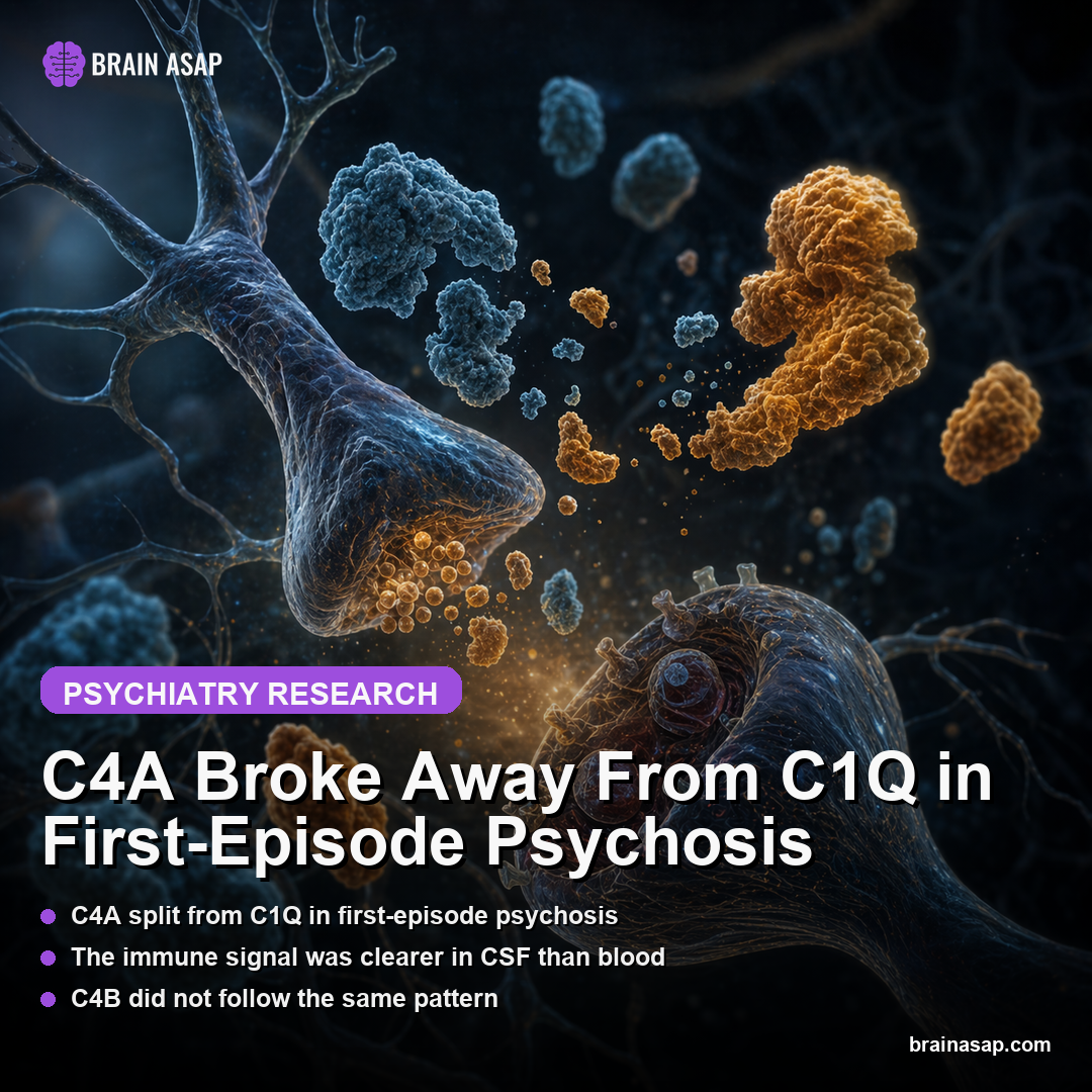
TL;DR: In cerebrospinal fluid from people with first-episode psychosis, the usual positive relationship between C4A and C1Q disappeared, while C4A’s broader immune-network pattern shifted sharply in a way C4B did not.
Key Findings
- C4A-C1Q coupling broke in psychosis: The study compared 113 patients with first-episode psychosis against 90 healthy controls drawn from the KaSP and GRIP cohorts.
- Healthy brains kept C4A and C1Q aligned: In controls, cerebrospinal fluid (CSF), the fluid surrounding the brain and spinal cord C4A and C1Q were positively associated at z = 0.41, p < 0.001, while C4B and C1Q were also positively linked.
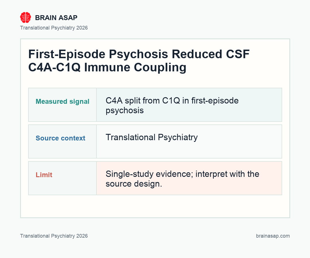
- That C4A-C1Q link vanished in psychosis: In first-episode psychosis, the CSF C1Q-C4A association dropped to z = 0.09, p = 0.40, suggesting selective decoupling rather than a blanket immune change.
- C4A shifted across 48 inflammatory proteins: Permutation testing showed a robust positive directional shift for C4A in psychosis at z = 3.81, p < 0.0001.
- C4B did not mirror C4A’s disease pattern: C4B showed a non-significant negative directional shift, reinforcing the idea that the two complement proteins are not interchangeable in psychosis biology.
- CSF and plasma told related but non-identical stories: The overall C4A shift broadly survived in plasma, yet individual protein-protein relationships differed sharply between compartments, arguing against a simple blood surrogate for the CSF signal.
Source: Translational Psychiatry (2026) | Arjmand et al.
For years, the complement model in schizophrenia has been summarized as “too much pruning.” This paper makes the mechanism both narrower and stranger: the important signal may not be the whole immune system rising together, but C4A behaving differently from its near-twin C4B inside the human CNS.
Why C4A Keeps Showing Up in Schizophrenia While C4B Usually Does Not
The complement hypothesis of schizophrenia got serious traction when genetic work pointed toward the major histocompatibility complex region and, more specifically, toward C4A copy number. That was intriguing because complement proteins are best known for immune defense, yet parts of the pathway also help tag synapses for pruning during development.
Ever since, the field has been stuck with a translation problem. If C4A really matters in schizophrenia risk, what exactly is it doing in living human brains, and how is that different from the closely related protein C4B? Genetic association by itself cannot answer that.
This study moves the question from DNA to fluid biology. By measuring complement proteins and a broad inflammation panel in both CSF and plasma, the authors ask whether C4A sits inside a distinct immune network in first-episode psychosis.
What 113 First-Episode Cases Revealed in Cerebrospinal Fluid
The cohort size is a strength here. The authors analyzed CSF from 113 patients with first-episode psychosis and 90 healthy controls, plus paired blood measurements and extensive inflammatory profiling. Importantly, 36 patients were antipsychotic-naive, which helps keep the whole result from collapsing into a medication artifact.
The first striking result was not that C4A was simply higher or lower. It was relational.
In healthy controls, C4A and C1Q moved together, just as you might expect if the classical complement pathway were operating in a coordinated way. In first-episode psychosis, that coordination disappeared.
- Controls: C4A and C1Q were positively linked in cerebrospinal fluid.
- First-episode psychosis: the C4A-C1Q relationship dropped away.
- C4B comparison: the near-twin protein did not reproduce the same disease pattern.
That is a more interesting kind of abnormality than a one-marker elevation. It implies that the network architecture around C4A may be altered, not just its absolute level.
How C4A, but Not C4B, Drifted Across the Inflammatory Network
The paper’s main claim becomes clearer when the authors expand beyond C1Q and look across 48 inflammation-related proteins. In healthy controls, C4A tended to show predominantly negative associations in CSF, while C4B and C1Q showed mostly positive ones. That already hints that C4A occupies a different niche.
In psychosis, the whole C4A pattern shifted. Using directional permutation tests, the authors found a robust positive shift for C4A at z = 3.81 with p < 0.0001.
C4B, by contrast, did not show the same disease-specific movement. That split is the point of the paper.
If you take schizophrenia’s complement findings seriously, this is the kind of evidence you want to see: not “inflammation happened,” but one candidate molecule behaving in a way that fits prior genetics and diverges from its closest biological look-alike.
CSF Still Looked More Informative Than Blood
The study also measured the same OLINK inflammatory markers in plasma, which matters because blood tests are much easier to deploy than lumbar punctures. The broad directional shift for C4A was still visible, but the underlying protein-by-protein map changed between plasma and CSF.
The warning is important. A blood marker can echo a central signal without reproducing the biology which makes the central signal interesting. Here the compartments were related enough to be encouraging, but different enough to show that plasma is not a plug-and-play substitute for brain-adjacent measurements.
The authors also checked whether a combined complement-plus-inflammatory profile tracked symptom severity. There was a hint that one CSF principal component related to negative symptoms, but it did not cleanly survive multiple-comparison correction. In other words, the strongest signal is still mechanistic, not yet clinical.
What the Selective C4A Signal Could Mean for Synaptic-Pruning Models
The most provocative interpretation is that C4A-driven synaptic tagging may become uncoupled from its usual complement context in early psychosis. The paper does not prove that directly, but it fits with a broader model in which C4A contributes to schizophrenia risk through a brain-specific immune specialization rather than a general inflammatory storm.
The reason is it pushes the field away from sloppy biomarker language. If C4A and C4B behave differently in living patients, then “complement activation” may be too blunt a phrase. The disease-relevant signal may sit in which complement relationships stay intact, which ones break, and in which compartment those breaks appear.
There are still limitations. The study is cross-sectional, the cohorts are clinically heterogeneous, and first-episode psychosis is not identical to schizophrenia.
Medication exposure is also part of the interpretation, even though the antipsychotic-naive subgroup helps. Early psychosis is biologically messy: stress, sleep disruption, immune state, substance exposure, and treatment timing can all shape inflammatory measurements. That is why the selective C4A pattern is interesting, but not yet diagnostic.
The next step is not simply to repeat the same measurement in a larger sample. The more useful test would follow people longitudinally from first episode into diagnostic clarification, symptom course, treatment exposure, and cognitive outcome, while measuring whether the C4A-C1Q relationship stays broken, normalizes, or marks a subgroup with a distinct trajectory.
That would also help answer the clinical translation question. A CSF immune-network signal is scientifically powerful, but lumbar puncture is not a routine screening tool.
If plasma can capture only the broad C4A shift while missing the central protein-map details, the field needs paired blood-CSF studies before treating blood complement markers as a shortcut.
But this is one of the better examples of a schizophrenia-linked immune finding becoming more specific rather than less specific as the data improve. That is usually a good sign the field is getting closer to the real mechanism.
Paper: Divergence of C4A and C4B in first-episode psychosis: Insights from CSF and plasma immune profiling. Translational Psychiatry. 2026;16:236.. DOI: 10.1038/s41398-026-04037-y
Authors: Arjmand et al.
Study Design: Cohort study
Sample Size: 203 people contributed CSF data: The study compared 113 patients with first-episode psychosis against 90 healthy controls drawn from the KaSP and GRIP cohorts.
Key Statistic: Healthy brains kept C4A and C1Q aligned: In controls, CSF C4A and C1Q were positively associated at z = 0.41, p < 0.001, while C4B and C1Q were also positively linked.