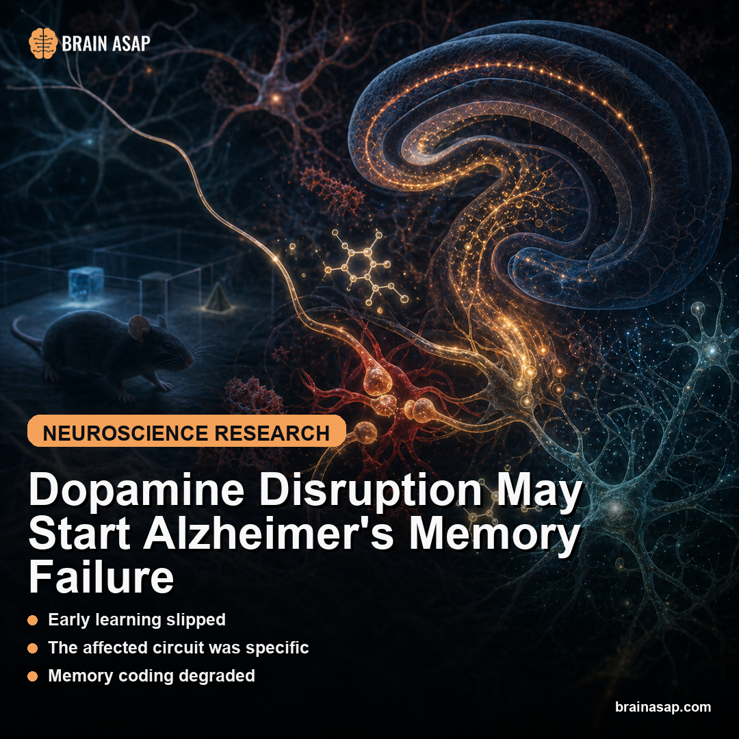
Dopamine Disruption May Start Alzheimer’s Memory Failure
TL;DR: In an Alzheimer’s mouse model, early memory failure tracked a broken dopamine signal into the lateral entorhinal cortex, and both optogenetic dopamine reactivation and L-DOPA partially restored learning.
Key Findings
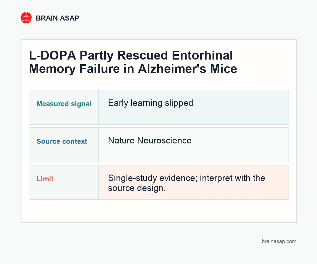
- Early learning slipped: Young APP knock-in mice reached 78.6% correct trials in the final learning block, compared with 90.6% in wild-type mice.
- The affected circuit was specific: Dopamine fibers from the ventral tegmental area and substantia nigra into the lateral entorhinal cortex lost task-related signaling during failed learning.
- Memory coding degraded: Layer 2/3 neurons in the lateral entorhinal cortex failed to keep odor and reward cues cleanly separated during learning.
- Direct circuit rescue worked: Optogenetic stimulation of lateral entorhinal dopamine fibers improved associative learning in APP knock-in mice.
- A drug rescue also worked: L-DOPA raised final-block performance to 82.6%, compared with 67.1% in saline-treated APP knock-in mice.
Source: Nature Neuroscience (2026) | Nakagawa et al.
Alzheimer’s disease is usually framed as an account of toxic protein buildup: amyloid plaques, tau tangles, and neurons slowly losing the ability to talk. This study points to a specific early failure. Before the memory system is simply gone, one of its teaching signals may stop arriving.
The Memory Gate That Fails Before the Door Closes
The entorhinal cortex is one of the first brain regions hit in Alzheimer’s disease. It acts like a gateway between sensory experience and the hippocampus, helping the brain decide which details belong together.
The new study focused on the lateral entorhinal cortex, a region that helps bind smells, cues, and outcomes into memory. In the task, mice learned which odor predicted reward and which odor predicted punishment. Healthy young mice learned the rule quickly; APP knock-in mice, which model amyloid biology, lagged behind.
The headline number is not subtle. By the final block of learning, wild-type mice were correct on 90.6% of trials, while the Alzheimer’s-model mice reached 78.6%. That gap appeared while the animals were still young, making it a candidate for an early circuit defect rather than a late-stage collapse.
APP knock-in mice are not designed to reproduce every feature of human Alzheimer’s disease, but they let researchers examine amyloid-linked circuit changes before broad neurodegeneration takes over. The model is useful for asking what breaks first, rather than only what is missing at the end.
- Wild-type mice: the healthy comparison group for normal odor-reward learning.
- APP knock-in mice: the amyloid-linked Alzheimer’s model used to test early circuit failure.
- L-DOPA-treated APP mice: the drug-rescue group used to test whether boosting dopamine could improve learning.
The task was also deliberately simple. A smell predicted reward or punishment, and the animal had to update its behavior as it learned the association. That simplicity is a strength: when performance falls, the researchers can look closely at the circuit that should be teaching the rule.
Dopamine Was the Teaching Signal
Dopamine is often described as a reward chemical, but that phrase undersells it. In memory circuits, dopamine helps mark what matters. It tells neural networks that a cue is worth updating, storing, and using later.
The team traced dopamine inputs from the ventral tegmental area and substantia nigra into the lateral entorhinal cortex. In healthy mice, those fibers carried learning-related signals when the animal correctly linked odor and outcome. In error sessions, that signal faded.
In APP knock-in mice, the same problem appeared earlier and more often. The dopamine fibers were still anatomically present, but their learning-related activity was unreliable.
The distinction is important: the circuit was not simply dead. It was under-signaling.
The authors also checked whether dopamine neurons in the midbrain were broadly lost. They did not find a simple disappearance of tyrosine hydroxylase-positive cells in the ventral tegmental area or substantia nigra. The more specific failure was functional: the learning signal reaching the lateral entorhinal cortex was not doing its job.
When Coding Gets Blurry
The researchers also recorded neurons in layer 2/3 of the lateral entorhinal cortex. In healthy mice, these cells learned to separate the odor cue from the outcome cue as training progressed. That separation is the neural version of understanding the rule.
In APP knock-in mice, the representation blurred. Odor and reward signals became less cleanly separated, which helps explain why behavior suffered. The animal was not just slower at the task; the relevant brain region was building a worse map of what the task meant.
This is the most interesting part of the paper. Amyloid was present, but the immediate failure looked like a dopamine-dependent encoding problem. That gives the study a circuit-level bridge between Alzheimer’s pathology and the everyday experience of memory confusion.
That bridge is important because memory is not a single storage bin. It is a set of operations: noticing the cue, assigning meaning, separating similar experiences, and using the right association later. The lateral entorhinal cortex sits inside that workflow, so a noisy teaching signal could create confusion before the system is structurally devastated.
Two Rescue Experiments Changed the Interpretation
The strongest evidence came from rescue experiments. When the researchers optogenetically reactivated lateral entorhinal dopamine fibers during the task, APP knock-in mice performed better. The intervention was targeted, timed, and anatomically specific.
Then the team tested L-DOPA, a dopamine precursor already used in Parkinson’s disease. L-DOPA-treated APP knock-in mice reached 82.6% correct trials in the final block, while saline-treated APP knock-in mice reached 67.1%. L-DOPA also helped restore the neural separation of cue representations.
The L-DOPA result belongs to mice, not clinical advice for people with Alzheimer’s. Timing, dose, disease stage, and dopamine biology do not translate cleanly from this experiment to patients, but the dopamine-entorhinal pathway now deserves attention as a possible early-stage mechanism.
The optogenetic experiment is especially persuasive because it avoided the bluntness of a whole-brain drug. The researchers stimulated the relevant dopamine fibers during the relevant task period. When that improved learning, it strengthened the argument that this pathway was causally involved rather than merely correlated with impairment.
Early Dopamine Failure Could Reframe Alzheimer’s Intervention Timing
Most Alzheimer’s drug development has aimed at amyloid, tau, inflammation, or broad neuroprotection. This paper suggests another layer: early failure in the modulatory signals that help memory circuits learn.
If a similar dopamine-entorhinal disruption exists in humans, it can help explain why memory problems can emerge before obvious large-scale tissue loss. It could also create a narrower target for imaging studies, pharmacology, or stimulation approaches.
The caveat is equally clear. This was a mouse model, not a human trial.
APP knock-in mice capture important amyloid-related biology, but they are not miniature Alzheimer’s patients. The study should be read as a strong mechanistic clue, not a treatment recommendation.
The next human-facing question is measurement. If entorhinal dopamine dysfunction contributes to early Alzheimer’s symptoms, researchers will need imaging, fluid biomarkers, or task-based physiology that can detect it in people. Without that bridge, the finding remains biologically exciting but clinically distant.
Even so, the paper widens the target. It suggests that early Alzheimer’s may involve not only toxic buildup inside vulnerable tissue, but also a breakdown in the modulatory signals that tell that tissue how to learn.
Entorhinal Dopamine Makes Alzheimer’s Look Less Amyloid-Only
The work reframes early Alzheimer’s memory failure as a problem of circuit instruction. The lateral entorhinal cortex can still be present, and its dopamine fibers can still be there, but the signal that teaches the circuit what to remember becomes unreliable.
That is a more hopeful kind of failure than irreversible destruction. A circuit that is mis-signaled may be harder to fix than a normal circuit, but easier to imagine rescuing than one that has already disappeared.
Paper: Early dopamine disruption in the entorhinal cortex of a knock-in model of Alzheimer's disease. Nature Neuroscience. 2026. DOI: 10.1038/s41593-026-02260-w
Authors: Nakagawa et al.
Study Design: Preclinical animal study
Sample Size: Early learning slipped: Young APP knock-in mice reached 78.6% correct trials in the final learning block, compared with 90.6% in wild-type mice.
Key Statistic: The affected circuit was specific: Dopamine fibers from the ventral tegmental area and substantia nigra into the lateral entorhinal cortex lost task-related signaling during failed learning.