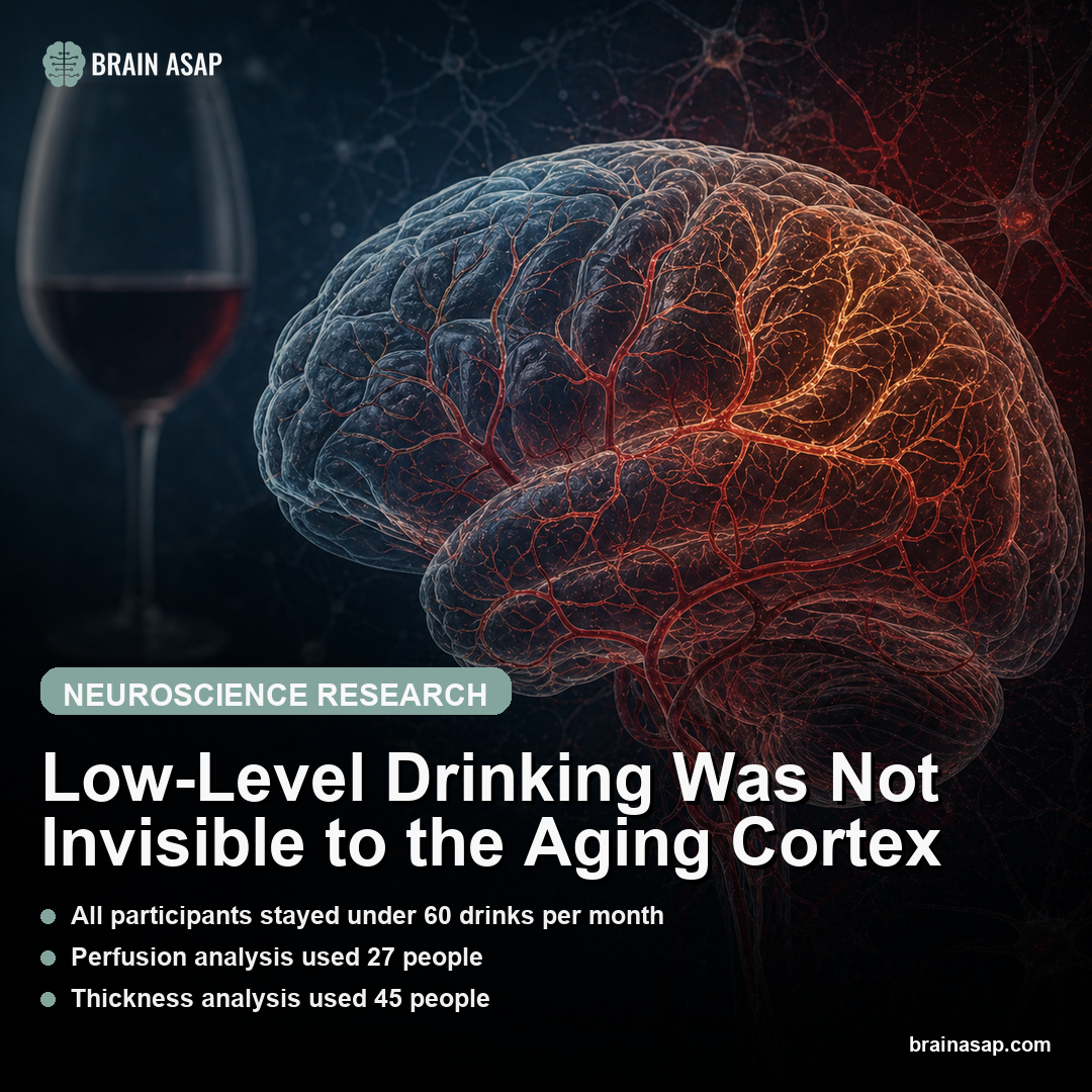
Low-Level Drinking Was Not Invisible to the Aging Cortex
TL;DR: Even drinking within low-risk guidelines was linked to lower cortical blood flow and thinner cortex when lifetime exposure met advancing age.
Key Findings
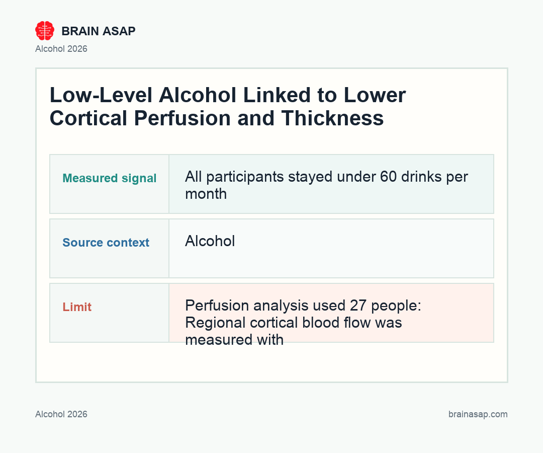
- All participants stayed under 60 drinks per month: The cohort excluded alcohol use disorder and focused on healthy non-smoking adults reporting low-level consumption.
- Perfusion analysis used 27 people: Regional cortical blood flow was measured with MRI in a smaller imaging subset.
- Thickness analysis used 45 people: Cortical morphometry tested whether lifetime drinking tracked with thinner cortex.
- Age multiplied the alcohol signal: The key predictor was age by total lifetime drinks, not a simple heavy-versus-light category.
- Frontal and parietal cortex stood out: Lower perfusion and thinner cortex appeared mainly in bilateral frontal, parietal, and related cortical regions.
Source: Alcohol (2026) | Mon et al.
Low-level alcohol consumption often gets treated as biologically quiet, especially when it stays inside guideline limits. This MRI study argues the aging brain may read the dose differently: not as a category of safe versus unsafe, but as a cumulative exposure that interacts with age.
Why Guideline-Level Drinking Is a Poor Brain Boundary
Public-health categories are useful, but the cortex does not necessarily honor them. A drink threshold designed around broad systemic risk may miss subtle tissue differences that accumulate over decades. This study deliberately stayed inside the low-level range, which makes the result more uncomfortable.
Participants were healthy, non-smoking adults aged 22 to 70 with no alcohol use disorder and no recent high-volume drinking. That design removes the easy explanation that the signal is just severe alcohol toxicity wearing a nicer label.
How Age Turned Lifetime Drinks Into an MRI Signal
The study’s central move was to treat alcohol exposure as continuous and cumulative. The average lifetime drinking level was about 21 drinks per month, and everyone reported no more than 60 standard drink equivalents per month in the previous year.
The important association emerged from the interaction between age and total lifetime drinks. Older adults with greater lifetime exposure showed lower cortical perfusion across multiple bilateral regions, with a broad average cortical perfusion signal rather than one isolated hot spot.
Blood Flow and Cortical Thickness Pointed to Vascular Aging
Perfusion is a live physiology measure: it asks how much blood is reaching cortical tissue. Thickness is a structural measure: it asks whether the tissue itself is slimmer. Seeing both move in the same broad direction makes the alcohol signal harder to dismiss as scanner noise.
The strongest regions were not random. Frontal and parietal cortices are central to executive function, attention, and cognitive aging. The paper does not prove low-level drinking causes decline, but it does place alcohol inside a brain-aging conversation that is usually reserved for heavier exposure.
Why Perfusion Is a Quiet but Important Brain-Aging Measure
Cortical perfusion is easy to overlook because it is less famous than gray matter volume or hippocampal size. But blood flow is one of the brain’s basic support systems. Neurons and glia depend on continuous oxygen and glucose delivery, and small vascular changes can shape cognition long before a scan shows obvious atrophy.
This is why the perfusion result is important. The study found lower regional cortical blood flow where the age-by-lifetime-drinking term was higher, including bilateral regions in frontal, parietal, and occipital cortex.
The signal was not framed as an acute intoxication effect. It looked more like a cumulative physiology association that becomes visible with aging.
Perfusion also sits at the intersection of alcohol and vascular aging. A small MRI study cannot untangle every route, but it can identify the cortical systems where the cumulative pattern appears:
- Vascular tone: alcohol can influence how blood vessels regulate flow.
- Sleep and blood pressure: both can mediate long-term brain perfusion risk.
- Inflammation: low-grade vascular stress could compound age-related cortical vulnerability.
Why Continuous Exposure Beats the Safe-or-Unsafe Drinking Box
The paper’s most useful modeling decision was to avoid treating low-level drinkers as one biologically identical group. Someone who drinks a few times per month for decades and someone who drinks near the guideline limit across adult life can both sit inside a low-risk category, yet their lifetime cortical exposure is not the same.
By using the interaction between age and total lifetime drinks, the authors tested a more realistic idea: the brain consequence of alcohol may depend on how long the tissue has been aging under that exposure. This is exactly the kind of relationship a categorical guideline can hide.
That does not show every drink is a measurable injury. It means the public-health boundary may be too blunt for brain tissue. The cortex may respond to dose history as a gradient, especially in systems already vulnerable to age-related vascular and structural change.
What This Study Can and Cannot Say About Guidelines
The sample size is the obvious limitation. 27 participants for perfusion and 45 for cortical thickness is enough to raise a mechanistic flag, not enough to rewrite national alcohol advice. The cohort was also cross-sectional, so lower perfusion and thinner cortex could reflect factors correlated with drinking rather than drinking itself.
Still, small mechanistic studies have a role. Large population datasets can tell us whether drinking predicts disease endpoints; MRI studies can suggest where in the brain a signal might begin. The ideal next step is a larger longitudinal cohort that measures lifetime alcohol exposure, vascular health, sleep, genetics, and repeated imaging together.
Until then, the practical interpretation is simple but not simplistic: low-level drinking should not be described as invisible to the brain. For people already thinking about dementia prevention, sleep quality, blood pressure, and vascular health, alcohol belongs in the same cumulative-risk conversation.
What a Larger Alcohol-Brain Study Should Measure Next
The next study needs repeated imaging. A single scan can show that people with different lifetime alcohol histories also differ in perfusion or thickness; longitudinal MRI can test whether those differences widen, stabilize, or reverse over time. That is the difference between a risk marker and a trajectory.
It also needs better context around sleep, blood pressure, sex, genetics, and drinking pattern. Two people can consume the same monthly total with very different timing: one drink every few days versus clustered weekend drinking. The brain may care about peaks, recovery time, and vascular stress as much as the average count.
Even with those gaps, the paper gives clinicians a more nuanced way to talk about alcohol. The question is not whether a patient meets criteria for alcohol use disorder. It is whether cumulative low-level exposure is one more modifiable factor in preserving cortical blood flow and structure with age.
Why the Result Lands Differently for Older Adults
The age interaction is the reason this paper feels different from a generic alcohol-health association. A 25-year-old and a 65-year-old may report similar low-level drinking, but the older cortex has accumulated more exposure while also facing vascular stiffening, sleep disruption, medication burden, and age-related tissue vulnerability.
That does not make alcohol uniquely dangerous compared with every other lifestyle factor. It makes it cumulative. The result asks readers to think less in terms of whether one drink is harmful and more in terms of whether decades of repeated exposure slightly tilt cortical physiology in the wrong direction.
Why a Small MRI Study Still Changes the Question
This is not a drinking guideline by itself. The perfusion sample was 27 people and the thickness sample was 45, so the result needs replication in larger cohorts with stronger confound control.
But the conceptual shift matters. The brain-health question is not only whether alcohol stays below a monthly limit. It is whether decades of exposure become more consequential as the cortex ages.
Paper: The interaction of age and total lifetime drinks is associated with regional cortical perfusion and thickness in healthy adults with low-level alcohol consumption. Alcohol. 2026. DOI: 10.1016/j.alcohol.2026.03.006
Authors: Mon et al.
Study Design: Cross-sectional MRI study of healthy non-smoking low-level alcohol consumers.
Sample Size: 27 participants for regional cortical perfusion and 45 for cortical volume/thickness analyses.
Key Statistic: Participants with greater age by lifetime drink exposure showed lower bilateral average cortical perfusion and lower bilateral average cortical thickness.