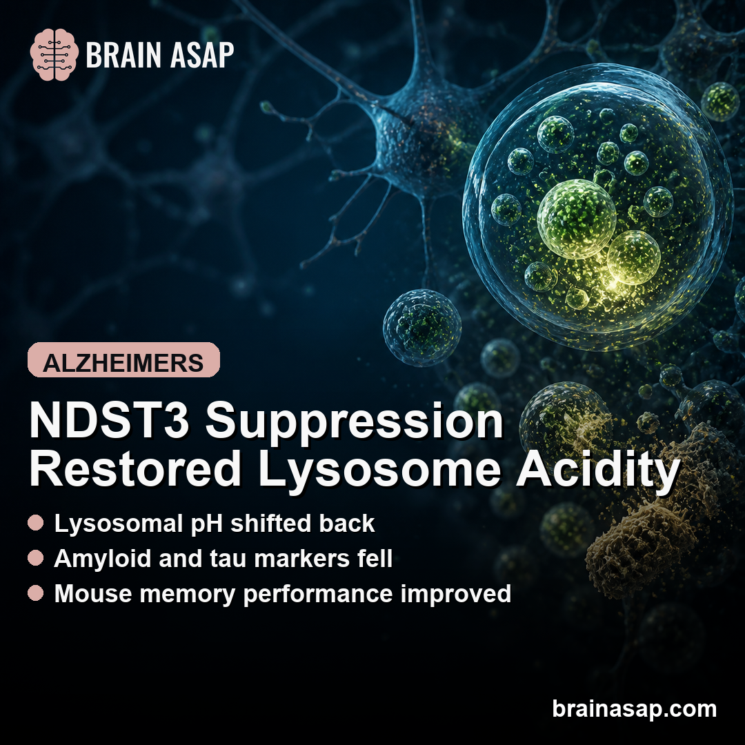
TL;DR: A 2026 study in Translational Neurodegeneration found that reducing NDST3, a microtubule deacetylase that affects lysosome acidity, restored lysosomal acidification and reduced amyloid-beta and tau pathology in Alzheimer’s cell and mouse models.
Key Findings
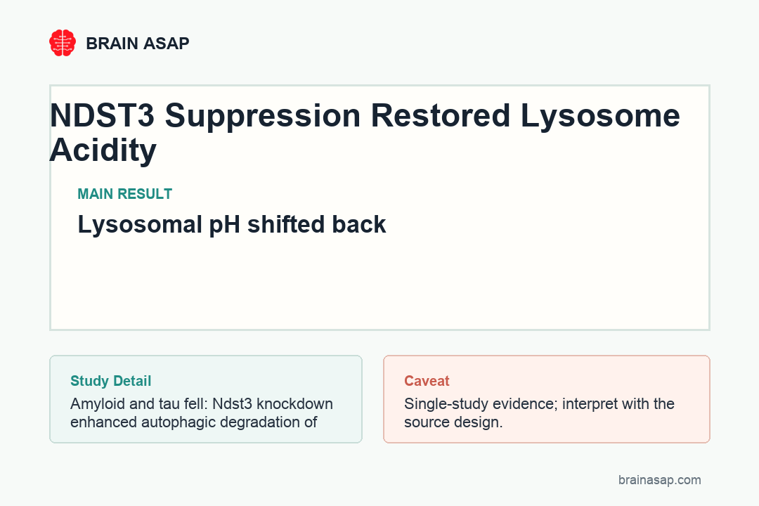
- Lysosomal pH shifted back: APP695Swe-overexpressing HT22 cells had lysosomal pH near 5.6, while Ndst3 knockdown lowered it to less than 5.0.
- V-ATPase assembly improved: NDST3 reduction restored lysosomal recruitment of V-ATPase V1 subunits, the machinery that helps acidify lysosomes.
- Amyloid and tau fell: Ndst3 knockdown enhanced autophagic degradation of amyloid-beta (Aβ), APP-CTFβ, and MAPT/tau in cell models.
- NDST3 was elevated in AD tissue: NDST3 expression was higher in APP695Swe cells, 3xTg-AD mouse hippocampus, and postmortem brain samples from Alzheimer’s patients.
- Mouse memory improved: 3xTg-AD mice with reduced NDST3 performed better in Morris water maze and novel object recognition tests than standard 3xTg-AD mice.
Source: Translational Neurodegeneration (2026) | Ge et al.
NDST3 is better known for heparan sulfate biology, but this study focused on a newer role: regulating microtubule acetylation and lysosomal acidification. Lysosomes need an acidic interior to degrade misfolded proteins, including Alzheimer’s-related amyloid-beta and tau species.
NDST3 Knockdown Reacidified Lysosomes in Alzheimer’s Cell Models
Researchers first compared NDST3 with HDAC6, another microtubule deacetylase already studied in Alzheimer’s disease. Both affected lysosomal pH, but NDST3’s activity was more concentrated around perinuclear microtubules, the region where highly acidic lysosomes are important for degradation.
In HT22 mouse hippocampal cells overexpressing APP695Swe, a mutant amyloid precursor protein used to model amyloid biology, lysosomal pH rose to about 5.6. That is less acidic than the roughly 4.7 pH seen under control conditions.
Reducing Ndst3 expression lowered lysosomal pH to less than 5.0. The mechanism involved better assembly of V-ATPase, the proton-pumping complex that acidifies lysosomes.
- V1 subunits returned: ATP6V1A and ATP6V1C1 levels increased in isolated lysosomal fractions after Ndst3 knockdown.
- V0 subunit stayed stable: ATP6V0D did not significantly differ across groups, suggesting improved assembly rather than a broad increase in all V-ATPase components.
- Perinuclear lysosomes recovered: LAMP2 staining suggested Ndst3 knockdown restored the perinuclear lysosome distribution needed for efficient autophagosome-lysosome fusion.
Autophagic Degradation of Aβ and MAPT/tau Increased
Restoring acidity mattered because lysosomal enzymes depend on pH. APP695Swe cells had lower cathepsin B activity and weaker dextran degradation, both signs of impaired lysosomal function.
After Ndst3 knockdown, cathepsin B activity increased and dextran degradation improved. The cells also showed signs of restored autophagic flux, including a better LC3B-II turnover response after chloroquine treatment.
The downstream Alzheimer’s markers moved in the expected direction:
- Aβ40 and Aβ42: Extracellular amyloid-beta levels were elevated in APP695Swe cells and decreased after Ndst3 knockdown.
- APP precursors: Full-length APP and APP-CTFβ were reduced after Ndst3 knockdown, and chloroquine blocked that reduction.
- Tau pathology: MAPT/tau P301L-overexpressing cells showed lower total tau and phosphorylated tau after Ndst3 knockdown, again with chloroquine blocking the effect.
NDST3 Was Higher in Alzheimer’s Models and Human Brain Samples
The researchers then checked whether NDST3 was actually altered in Alzheimer’s-related tissue. It was.
NDST3 mRNA and protein were higher in APP695Swe-overexpressing HT22 cells. In 3xTg-AD mice, NDST3 protein was elevated across hippocampal CA1, CA2, CA3, and dentate gyrus regions at 3, 6, and 12 months.
Postmortem human brain samples from Alzheimer’s patients also showed higher NDST3 staining than healthy control samples. The human sample was small, with 3 subjects per group, but it supported the same direction seen in the experimental models.
Reduced NDST3 Lowered Amyloid Plaques and p-tau in 3xTg-AD Mice
For the in vivo test, researchers bred 3xTg-AD mice with partial Ndst3 knockout mice. The resulting 3xTg-Ndst3+/- mice had reduced NDST3 expression in the hippocampus.
At 10 months, standard 3xTg-AD mice showed more poorly acidified autolysosomes, higher P62 accumulation, and altered LC3B-II/I ratio. Partial NDST3 reduction reversed those lysosomal/autophagy markers toward the nontransgenic control pattern.
Alzheimer’s pathology also decreased. Compared with 3xTg-AD littermates, 3xTg-Ndst3+/- mice had lower hippocampal APP-CTFβ and MAPT/tau levels, fewer amyloid plaques, and lower phosphorylated MAPT/tau Ser422 immunoreactivity.
- Neuron structure: Nissl, Fluoro-Jade C, NeuN, Golgi, and electron microscopy measures pointed toward less neuronal impairment after NDST3 reduction.
- Microglia: Iba1 and CD68/Iba1 signals suggested lower microglial load and activation.
- Astrocytes: GFAP area, reduced in the 3xTg-AD model, was partly restored after NDST3 reduction.
Water Maze and Novel Object Memory Improved After NDST3 Reduction
Cognitive testing used 12 mice per group. Swimming speed did not differ before Morris water maze testing, which helped reduce the chance that motor performance explained the memory results.
Standard 3xTg-AD mice took longer paths and longer time to find the hidden platform. Mice with partial NDST3 reduction showed shorter escape latencies and swimming distances than 3xTg-AD controls.
During the probe test, 3xTg-Ndst3+/- mice spent more time in the target quadrant and crossed the former platform location more often. Novel object recognition showed a similar pattern: the partial-knockout mice, unlike standard 3xTg-AD mice, preferred the novel object during testing.
NDST3 Is a Target Hypothesis, Not an Alzheimer’s Treatment Yet
The results support NDST3 as a mechanistic target for restoring lysosomal acidification in Alzheimer’s models. They do not show that an NDST3 drug is ready for human testing.
Several limits keep the conclusion preclinical. The researchers used cell overexpression models and 3xTg-AD mice, not patients receiving a treatment.
The human brain comparison had only 3 Alzheimer’s cases and 3 controls. The study also used genetic NDST3 reduction rather than a selective NDST3 inhibitor.
- Drug gap: No pharmacological NDST3 blocker was tested.
- Model gap: The APP695Swe cell system overexpresses mutant APP while endogenous APP remains present.
- Timing gap: The mouse work did not establish a long longitudinal treatment window for progressive cognitive decline.
The strongest claim is mechanistic: in Alzheimer’s models with under-acidified lysosomes, reducing NDST3 restored lysosomal function and lowered several downstream pathology markers. Turning that mechanism into a therapy would require a drug, safety work, and validation in models closer to human Alzheimer’s disease.
Citation: DOI: 10.1186/s40035-026-00549-1. Ge et al. NDST3 suppression restores lysosomal acidification and ameliorates amyloid-β and MAPT/tau pathology in Alzheimer’s disease. Translational Neurodegeneration. 2026;15:16.
Study Design: Cell and mouse mechanism study using HT22 Alzheimer’s-related overexpression models, postmortem human brain tissue, and 3xTg-AD mice with partial Ndst3 knockout.
Sample/Model: HT22 mouse hippocampal cells, 3xTg-AD mice, 12 mice per behavioral group, and postmortem human brain samples from 3 Alzheimer’s patients and 3 controls.
Key Statistic: APP695Swe cells had lysosomal pH near 5.6, and Ndst3 knockdown lowered it to less than 5.0 while reducing amyloid/tau markers and improving memory-test performance in 3xTg-AD mice.
Caveat: The work is preclinical and genetic; no selective NDST3 drug was tested in humans or animals.