TL;DR: A 2026 article-in-press study in Alzheimer’s Research & Therapy found that olfactory-region MRI radiomics, a method that turns brain scans into quantitative texture and shape features, linked blood pTau217, a tau-related Alzheimer’s disease biomarker, with cognitive impairment across three cohorts.
Key Findings
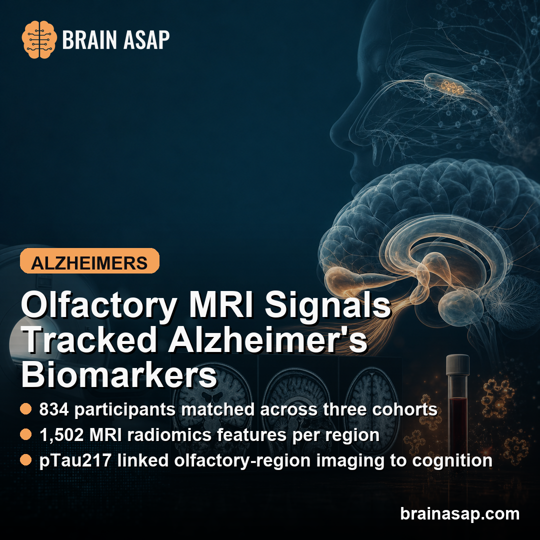
- Three Alzheimer’s cohorts were matched: Researchers matched 122 Alzheimer’s disease (AD) patients and 156 cognitively unimpaired (CU) controls in each cohort, for 834 participants total.
- 1,502 MRI radiomics features per region: Structural MRI scans were converted into quantitative shape, intensity, and texture features from bilateral olfactory-related brain regions.
- Hippocampus-amygdala features performed best: The strongest models reached AUC 0.86-0.92, where AUC summarizes how well a model separates AD from CU participants.
- Radiomics improved sensitivity: Compared with conventional hippocampus-amygdala volume models, radiomics showed stronger AD detection sensitivity (0.776-0.869 versus 0.505-0.811) while AUC was similar.
- pTau217 connected to cognition through MRI texture: Two hippocampal radiomics features statistically mediated 26.9%-37.1% of the relationship between plasma pTau217 and Mini-Mental State Examination (MMSE), a cognitive screening test.
Source: Alzheimer’s Research & Therapy (2026) | Chen et al.
pTau217 is phosphorylated tau 217, a blood marker tied to Alzheimer’s tau pathology. It can help identify disease biology, but a blood value does not show where tissue-level brain changes are happening.
Radiomics tries to fill part of that spatial gap. Instead of measuring only whether a hippocampus is smaller, radiomics extracts many quantitative features from MRI images, including intensity patterns and texture variation that may reflect microstructural change.
Olfactory Brain Regions Were Tested Across Three Alzheimer’s Cohorts
Researchers focused on smell-related brain systems because olfactory impairment often appears early in Alzheimer’s disease and overlaps with medial temporal regions involved in memory. The analysis covered the hippocampus, amygdala, piriform cortex, entorhinal cortex, and orbitofrontal cortex.
The study used three cohorts with the same AD-versus-CU counts in each dataset: 122 AD patients and 156 CU controls. The in-house cohort included olfactory testing, plasma pTau217, and a broader cognitive battery.
ADNI and OASIS served as external imaging-validation cohorts.
- Clinical groups: AD patients had lower MMSE scores and higher Clinical Dementia Rating scores than CU controls across cohorts.
- Olfactory testing: In the in-house cohort, AD patients had lower odor-identification scores (6.45 versus 10.50).
- Blood biomarker: In the same cohort, AD patients had higher plasma pTau217 (0.80 versus 0.15 pg/mL).
Because the MRI scans came from different scanners and sites, researchers used ComBat harmonization to reduce scanner-related batch effects before training the models. The harmonization step is important because radiomics can be sensitive to technical differences that are not disease biology.
Hippocampus-Amygdala Radiomics Classified Alzheimer’s Best
Across the five olfactory-related region sets, hippocampus-amygdala radiomics produced the most stable AD classification. Six machine-learning algorithms were tested: support vector machine, logistic regression, random forest, LightGBM, Gaussian naive Bayes, and XGBoost.
Logistic regression and support vector machine performed best. In external validation, both reached AUC values of 0.883 in ADNI and 0.922 in OASIS.
Logistic regression had slightly higher sensitivity, while support vector machine had higher specificity.
The model comparison also separated radiomics from a simple hippocampal-volume finding. Radiomics and volume models had similar average AUC values, 0.872 and 0.868, but radiomics showed stronger sensitivity for detecting AD cases.
- Radiomics sensitivity: Across cohorts and algorithms, sensitivity ranged from 0.776 to 0.869.
- Volume-model sensitivity: Comparable volume models ranged from 0.505 to 0.811.
- Cognitive prediction: Radiomics features added significant explanatory value in 75.7% of all-participant analyses, compared with 12.5% for volumetric features.
pTau217 and MMSE Were Linked Through Hippocampal Texture Features
The in-house cohort allowed researchers to connect the MRI features with plasma pTau217, smell testing, and cognition. The hippocampus-amygdala radiomics score correlated with several clinical readouts across all participants.
Higher radiomics scores were associated with several clinical measures after false-discovery-rate correction:
- Global cognition: MMSE correlation r = -0.65.
- Olfactory identification: Smell-test correlation r = -0.48.
- Language and speed: Verbal fluency correlation r = -0.45; SDMT processing-speed correlation r = -0.49.
- Blood biomarker: Plasma pTau217 correlation r = 0.49.
Within AD patients alone, the radiomics score still correlated with MMSE (r = -0.28). In CU controls, the correlations were minimal, which suggests the signature was more tied to disease-related change than ordinary aging in this dataset.
Mediation Analyses Connected Tau, Smell, and Cognitive Scores
The mediation analyses asked whether specific MRI texture features statistically sat between the biomarker or smell measure and cognitive performance. These models cannot prove causality, but they are useful for testing whether a brain-imaging feature helps account for a clinical association.
- pTau217 to MMSE: A left hippocampal radiomics feature mediated 37.1% of the plasma pTau217-MMSE relationship.
- Second hippocampal feature: A right hippocampal texture feature mediated 26.9% of the same pTau217-MMSE relationship.
- Odor identification to cognition: The hippocampus-amygdala radiomics score mediated 26.6% of the odor-language relationship and 44.2% of the odor-memory relationship.
Read plainly, the paper supports a regional pathway: tau-related blood biomarker burden and olfactory dysfunction were associated with hippocampus-amygdala MRI texture features, and those features were associated with worse cognitive performance.
The Main Limits Are Cross-Sectional Design and Biomarker Scope
The most important caution is that the design was cross-sectional. The study can show linked measurements at one point in time, but it cannot prove that pTau217 caused the MRI texture changes or that those features predicted future decline.
Several other constraints keep the result in biomarker-development territory rather than ready clinical screening:
- Clinical AD classification: AD diagnosis relied on NINCDS-ADRDA clinical criteria without amyloid positron emission tomography (PET) or cerebrospinal-fluid confirmation.
- Limited multimodal validation: pTau217 and olfactory analyses were limited to the in-house cohort, not replicated with matching biomarker and smell data in ADNI or OASIS.
- Biological interpretation: Radiomics features need post-mortem or molecular-imaging validation before researchers can say exactly what tissue changes each feature represents.
- Prodromal disease: The study did not test whether these signatures predict conversion from mild cognitive impairment to Alzheimer’s dementia.
Even with those limits, the study gives a concrete imaging target for future work: hippocampus-amygdala radiomics may add regional information to blood pTau217 and smell testing when researchers are trying to map Alzheimer’s biology onto cognition.
Citation: DOI: 10.1186/s13195-026-02053-0. Chen et al. Olfactory radiomics signatures link pTau217 to cognitive impairment in probable Alzheimer’s disease: a multi-cohort machine learning study. Alzheimer’s Research & Therapy. 2026.
Study Design: Multi-cohort structural MRI radiomics and machine-learning study with biomarker, olfactory, and cognitive analyses in the in-house cohort.
Sample Size: 834 participants across three cohorts: 366 AD patients and 468 CU controls.
Key Statistic: Hippocampus-amygdala radiomics models reached AUC 0.86-0.92, and two hippocampal radiomics features mediated 26.9%-37.1% of the plasma pTau217-MMSE relationship.
Caveat: The design was cross-sectional, AD classification was clinical rather than amyloid-confirmed, and mechanistic pTau217 analyses were limited to one cohort.