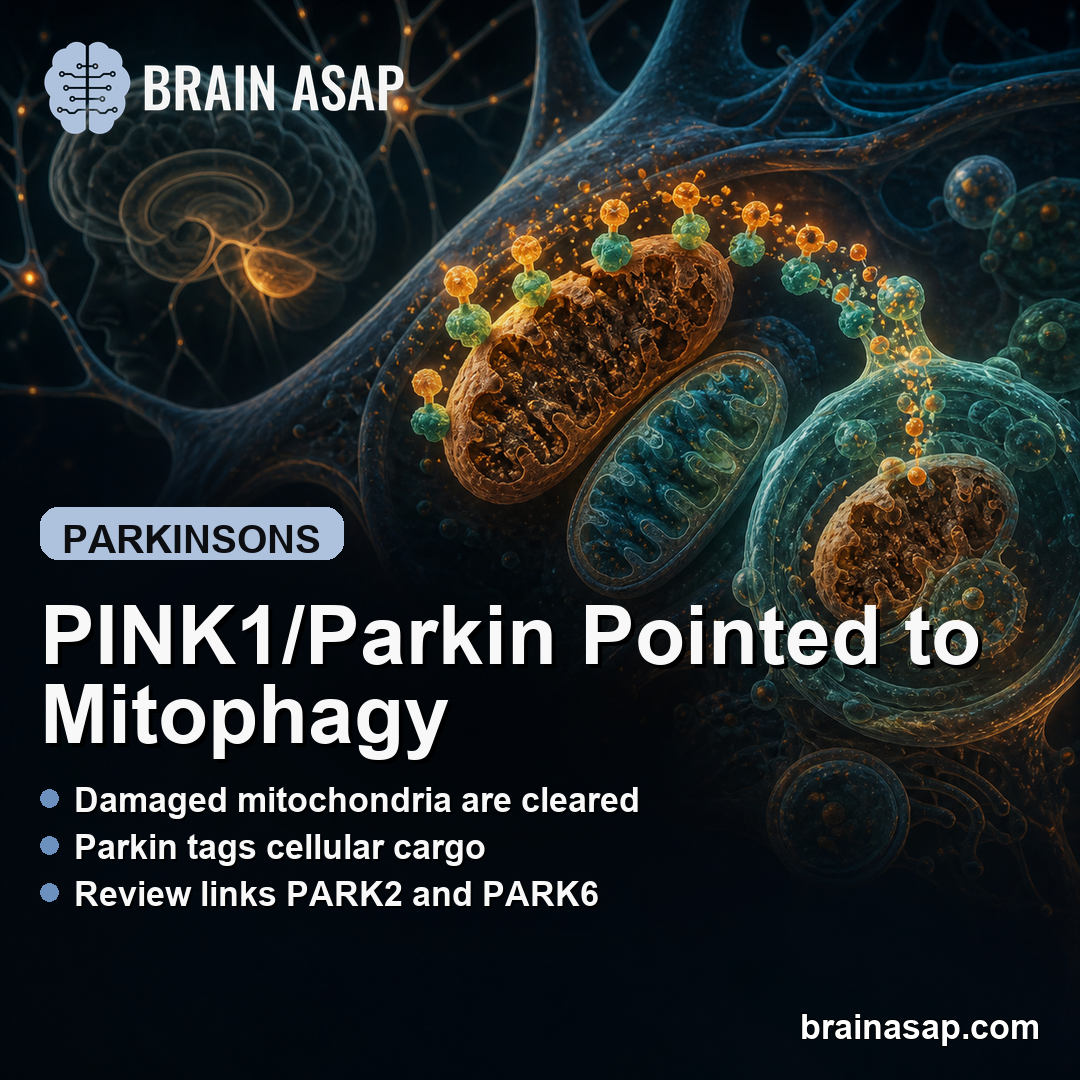
TL;DR: A 2026 review in Translational Neuroscience summarizes why PINK1 and parkin, two genes linked to early-onset parkinsonism, keep pointing researchers back to mitophagy and autophagy as Parkinson disease mechanisms.
Key Findings
- 18 PARK loci noted: The review frames familial Parkinson genetics as a route into molecular mechanisms, including PARK2 and PARK6.
- PARK2 encodes parkin: Parkin is an E3 ubiquitin ligase linked to autosomal recessive parkinsonism.
- PARK6 encodes PINK1: PINK1 is a kinase that accumulates on damaged mitochondria and helps recruit parkin.
- Mitophagy is central: The review emphasizes removal of damaged mitochondria as a major PINK1/parkin pathway.
- MIRO1 and ROCK2 matter: Recent studies highlighted MIRO1 as a parkin docking factor and ROCK2 inhibition as a possible mitophagy-stimulating route.
Source: Translational Neuroscience (2026) | Stegmuller
PINK1 and Parkin Keep Parkinson Disease Focused on Mitochondria
PINK1/parkin-associated mitophagy is the review’s main thread. The review does not claim that every Parkinson disease case starts with these genes; it explains why these genes have become a useful route into the cell biology of parkinsonism.
Parkinson disease involves loss of dopaminergic neurons, motor symptoms such as resting tremor and bradykinesia, and non-motor symptoms that include depression, sleep disturbance, fatigue, autonomic dysfunction, and cognitive deficits.
The review notes that Parkinson cases nearly tripled between 2010 and 2023 in a meta-analysis covering more than 40 years of patient data. That growing burden makes mechanism work more than a genetics exercise.
- PARK2: This locus encodes parkin, an E3 ubiquitin ligase that marks proteins for downstream handling.
- PARK6: This locus encodes PINK1, a kinase involved in detecting mitochondrial damage.
- Early-onset phenotype: Mutations in these genes are tied to autosomal recessive parkinsonism, often with earlier onset and slower progression.
- Cell-cleanup pathway: Their best-known shared pathway is mitophagy, the selective autophagy of damaged mitochondria.
The practical reason to care is that mitochondria are not passive energy units in neurons. Damaged mitochondria can disturb energy supply, calcium handling, stress signaling, and protein quality control.
Dopaminergic neurons are especially sensitive to those stresses because they have long axons, high energy demand, and continuous synaptic signaling requirements. A small defect in mitochondrial maintenance can therefore become a larger problem over years.
The review uses familial Parkinson genes as a mechanistic entry point. That does not make PINK1 and parkin the only relevant proteins, but it gives researchers a concrete pathway to test rather than treating Parkinson disease as one undifferentiated process.
Mitophagy Removes Damaged Mitochondria Before They Accumulate
Autophagy is the broader cell-cleanup system that packages cellular material for lysosomal degradation. Mitophagy is the mitochondrial version of that process.
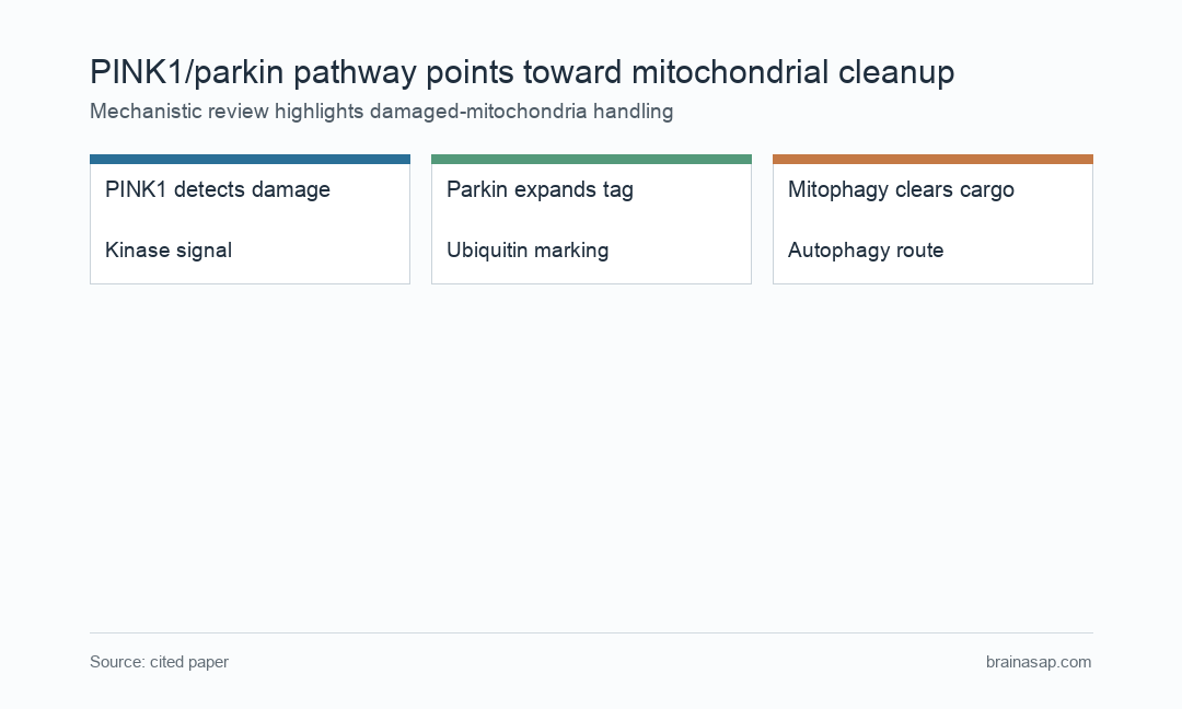
In the PINK1/parkin model, mitochondrial damage changes the surface of the organelle. PINK1 accumulates there, marks the damaged mitochondrion through phosphorylation events, and helps recruit parkin to expand the ubiquitin signal.
- Damage recognition: PINK1 becomes stabilized on dysfunctional mitochondria instead of being cleared quickly.
- Signal amplification: Parkin adds ubiquitin tags that help label mitochondrial proteins for removal.
- Autophagy engagement: Autophagy machinery can then isolate and deliver damaged mitochondrial material toward lysosomal degradation.
MIRO1 and ROCK2 Add New Detail to the Cleanup Pathway
The review highlights recent work on MIRO1, a mitochondrial outer-membrane protein involved in mitochondrial movement. Several studies suggest MIRO1 helps position parkin at damaged mitochondria.
Neurons depend on mitochondrial transport along long processes. If docking, movement, and cleanup are linked, then mitochondrial quality control is also a wiring-maintenance problem.
- MIRO1 docking: A subset of parkin can interact with MIRO1 before mitochondrial damage becomes obvious.
- Damage response: PINK1 accumulation after damage can activate parkin and promote MIRO1 ubiquitination.
- Delayed clearance: Loss of MIRO1/2 was reported to delay parkin translocation and impair removal of dysfunctional mitochondria in neurons.
- ROCK2 inhibition: The review also describes work in which ROCK inhibitors, especially SR3677 against ROCK2, stimulated parkin-mediated mitophagy steps.
These examples show why the pathway is still being revised. PINK1 and parkin are central, but accessory proteins and kinase pathways may decide where, when, and how efficiently mitophagy occurs.
Another important point is that mitophagy is not an isolated switch. It intersects with mitochondrial transport, lysosomal function, stress responses, and protein turnover, which are all relevant to neuronal survival.
That wider network is why a simple “increase autophagy” approach may be too blunt. The more useful question is whether a specific intervention restores damaged-mitochondria handling without disrupting normal cellular recycling.
The review therefore treats mitophagy as a pathway to map carefully, not a slogan. The same cleanup machinery that protects neurons must still preserve healthy mitochondria and normal cellular balance.
Genetic Parkinson Mechanisms Do Not Equal a Ready Treatment
The review is mechanistic. It collects cell, animal, and molecular evidence around a pathway, but it does not prove that boosting mitophagy will slow Parkinson disease in patients.
That distinction is important because familial PINK1/parkin parkinsonism is not the same as typical late-onset Parkinson disease. A therapy that helps one biological subgroup may not generalize to every patient.
- Translation gap: Mitophagy markers in cells do not automatically predict clinical benefit.
- Disease timing: Neuronal damage may begin long before a diagnosis, so rescue windows may be early and narrow.
- Pathway complexity: Autophagy can be protective or harmful depending on context, dose, and disease stage.
PINK1 and parkin keep Parkinson research focused on mitochondrial quality control. The pathway gives researchers specific proteins to test, but patient-facing treatment claims still need clinical evidence.
The review helps explain why Parkinson genetics and mitochondrial biology are often discussed together. The strongest evidence is still mechanistic, but the pathway gives drug discovery a more testable set of targets than broad neuroprotection language.
Citation: DOI: 10.1515/tnsci-2025-0386. Stegmuller. Novel insight into PINK1/parkin-associated autophagy implicated in Parkinson disease. Translational Neuroscience. 2026.
Study Design: Narrative mechanistic review of PINK1/parkin-associated autophagy and mitophagy in Parkinson disease biology.
Sample/Model: Review of genetic, cell-biological, animal-model, and molecular studies rather than a new patient cohort.
Key Statistic: The review highlights PARK2/parkin and PARK6/PINK1 among 18 PARK loci and centers mitophagy as a major pathway.
Caveat: Mechanistic review evidence does not prove that mitophagy-targeting interventions improve patient outcomes.