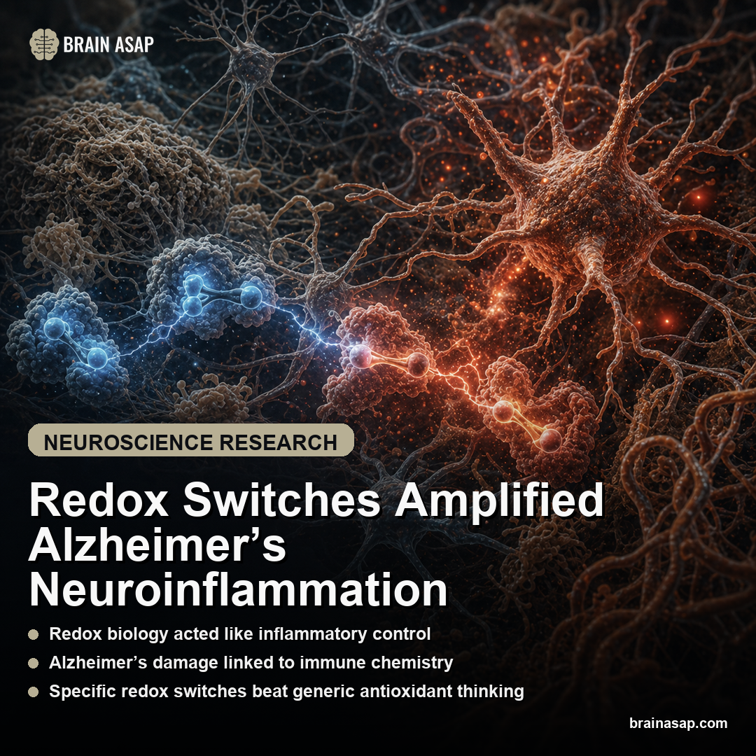
TL;DR: A 2026 study in Cell Chemical Biology found that alzheimer’s inflammation may be driven by a specific chemical switch: S-nitrosylation of STING at cysteine 148, which pushed innate immune signaling toward synaptic damage.
Key Findings
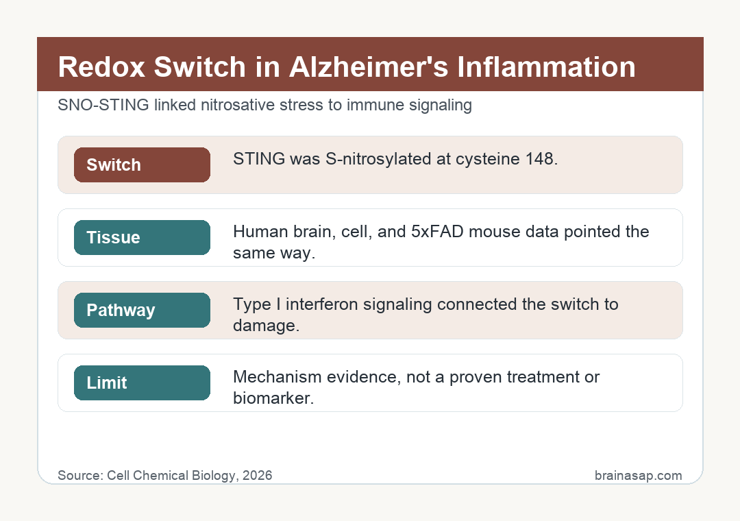
- STING carried the redox switch: The study identified S-nitrosylation of human STING at cysteine 148 as a redox modification tied to Alzheimer’s inflammatory signaling.
- Human Alzheimer’s brain showed SNO-STING: The redox-modified STING form was observed in postmortem Alzheimer’s disease brain tissue.
- Three disease-relevant systems converged: The readout appeared in human AD brain, hiPSC-derived innate immune cells exposed to amyloid-beta and alpha-synuclein aggregates, and 5xFAD transgenic mice.
- Oligomerization explained the inflammatory push: S-nitrosylation promoted STING oligomer formation, which can drive type I interferon signaling.
- The treatment target narrowed: The therapeutic target is not generic antioxidant cleanup but a redox-sensitive cysteine that may control pathological STING activation.
Source: Cell Chemical Biology (2026) | Carnevale et al.
Oxidative stress is often treated as background damage in Alzheimer’s disease, like cellular rust accumulating around amyloid and tau. This Cell Chemical Biology paper makes the chemistry more active.
It argues that nitrosative stress can flip a specific immune switch inside the cGAS-STING pathway, turning a stress readout into excessive brain inflammation.
STING Linked Nitrosative Stress to Alzheimer’s Inflammation
The key protein is STING, short for stimulator of interferon genes.
Study details:
- STING carried the redox switch: The study identified S-nitrosylation of human STING at cysteine 148 as a redox modification tied to Alzheimer’s inflammatory signaling
- Human Alzheimer’s brain showed SNO-STING: The redox-modified STING form was observed in postmortem Alzheimer’s disease brain tissue
- Three disease-relevant systems converged: The readout appeared in human AD brain, hiPSC-derived innate immune cells exposed to amyloid-beta and alpha-synuclein aggregates, and 5xFAD transgenic mice
- Oligomerization explained the inflammatory push: S-nitrosylation promoted STING oligomer formation, which can drive type I interferon signaling
STING is part of the innate immune system: it helps cells respond when DNA appears in the wrong compartment, a danger readout that can happen during infection, mitochondrial stress, or tissue damage.
That defense system becomes risky in neurodegeneration.
If STING signaling stays too active, it can drive type I interferon programs, inflammasome activity, and other inflammatory cascades that make vulnerable brain tissue worse rather than better.
The study’s central move was to connect that immune pathway to S-nitrosylation, a chemical modification in which nitric-oxide-related species attach to a cysteine residue on a protein.
In this case, the residue was cysteine 148 on human STING. Proteins are not only controlled by how much of them a cell makes.
They are also controlled by small chemical modifications that change shape, binding, location, or signaling strength. S-nitrosylation is one of those modifications.
Here, cysteine 148 mattered because it helped STING form oligomers, clusters of STING molecules that create the active platform for downstream signaling.
When STING oligomerizes, it can recruit TBK1 and IRF3 signaling, pushing cells toward interferon-stimulated gene expression.
That is why the paper is more specific than a general oxidative-stress claim.
The mechanism is not “oxidants are bad.” It is nitrosative chemistry modified a defined cysteine on a defined immune protein, and that modification promoted inflammatory signaling relevant to Alzheimer’s disease.
Human Brain, Cell Models, and 5xFAD Mice Pointed the Same Way
The evidence is strongest because the mechanism did not sit in one isolated assay. Researchers reported S-nitrosylated STING, or SNO-STING, in postmortem human Alzheimer’s disease brain tissue.
They also tested human induced pluripotent stem cell-derived innate immune cells exposed to Alzheimer’s-related aggregates, including amyloid-beta and alpha-synuclein.
Those cell models matter because they let researchers expose human immune-like cells to disease-relevant protein stress while controlling the system more tightly than a whole brain allows.
- Human Alzheimer’s brain tissue: showed the redox-modified STING readout in postmortem samples.
- Human cell-derived immune models: let researchers expose innate immune cells to amyloid-beta and alpha-synuclein aggregates under controlled conditions.
- 5xFAD mice: tested the same pathway in an amyloid-heavy Alzheimer’s model with neuroinflammatory changes.
The third line of evidence came from 5xFAD mice, a transgenic Alzheimer’s model that develops amyloid pathology and inflammatory changes.
No mouse model captures human Alzheimer’s disease fully, but convergence across human tissue, human cell-derived systems, and an AD mouse model makes the redox-STING link more credible as a one-platform artifact.
Microglial Interferon Signaling Is the Damage Pathway
Microglia are the brain’s resident immune cells.
They can clear debris and respond to injury, but when inflammatory programs become chronic or excessive, they can contribute to synaptic loss and neuronal stress.
The STING pathway is relevant because it sits upstream of type I interferon signaling.
Interferons are helpful antiviral molecules, but in the wrong context they can help sustain inflammatory damage.
In Alzheimer’s disease, the distinction is important because the brain is not fighting a simple infection; it is responding to accumulating protein aggregates, mitochondrial stress, and damaged tissue.
The study suggests SNO-STING helps push that immune pathway into a pathological state.
In the 5xFAD model, researchers connected the signaling cascade to early neuroinflammation and synaptic loss, which is closer to the clinical problem than inflammation measured for its own sake.
The synapse angle is the reason the mechanism matters for cognition.
Alzheimer’s symptoms track poorly with plaque count alone, but synaptic loss is much closer to memory failure.
A redox switch that pushes microglia and innate immune signaling toward synaptic damage therefore sits closer to the disease process patients and families actually experience.
Targeting SNO-STING Is Different From Taking Antioxidants
The treatment implication is precision, not supplement hype. Alzheimer’s trials have repeatedly shown that broad biological plausibility does not guarantee clinical benefit, and generic antioxidant logic is especially easy to overstate.
This paper points to a narrower idea: if cysteine 148 helps pathological STING activation, then a therapy might eventually target that redox-sensitive site or the S-nitrosylation reaction around it.
The goal would be to reduce damaging neuroinflammation while preserving the immune functions STING normally performs.
That last part is crucial.
STING is part of innate immune defense, so a blunt shutdown could create its own problems.
The attractive idea is selective modulation of pathological SNO-STING signaling, not wiping out the pathway entirely.
That also explains why the cysteine 148 result is more than a molecular footnote.
A drug target becomes more plausible when researchers can point to a specific residue, a specific modification, and a specific downstream pathway.
It gives medicinal chemistry a smaller target than “inflammation,” which is too broad to be helpful by itself.
The Alzheimer’s Claim Is Mechanistic, Not Clinical Yet
The evidence is mechanistic rather than clinical. Blocking SNO-STING has not been shown to slow cognitive decline in people, and redox biology is not yet a ready biomarker.
What it does provide is a molecular bridge from nitrosative stress to immune activation to synaptic damage.
That bridge is valuable because Alzheimer’s disease is often discussed as if amyloid and tau are the whole map.
They are central, but tissue injury also depends on how brain cells and immune cells respond to them.
The mechanistic implication is that Alzheimer’s inflammation may be chemically switched at the protein level.
If that switch can be targeted without disabling normal immunity, it gives researchers a more concrete therapeutic route than “reduce oxidative stress” ever did.
The next experiments should test whether blocking STING S-nitrosylation preserves synapses, changes microglial state, and improves behavior in disease models without weakening helpful immune defense.
Those are the steps needed before SNO-STING becomes a credible therapeutic program rather than an elegant mechanism, especially for a disease where many plausible targets have failed after moving into harder tests.
Citation: DOI: 10.1016/j.chembiol.2026.03.017. Carnevale et al. Redox regulation of neuroinflammatory pathways contributes to damage in Alzheimer's disease brain. Cell Chemical Biology. 2026
Study Design: Redox chemical-biology and mass-spectrometry study using human Alzheimer’s postmortem brain tissue, hiPSC-derived innate immune cells, and 5xFAD transgenic mice.
Sample/Model: Human postmortem Alzheimer’s brain tissue, human cell-derived innate immune models, and 5xFAD mouse experiments; exact group counts should be checked in the full paper before publication.
Key Statistic: The central molecular result was S-nitrosylation of human STING at cysteine 148, a modification linked to STING oligomerization and excessive type I interferon signaling.
Caveat: The study identifies a disease mechanism and therapeutic target, not a proven Alzheimer’s treatment or clinical biomarker.