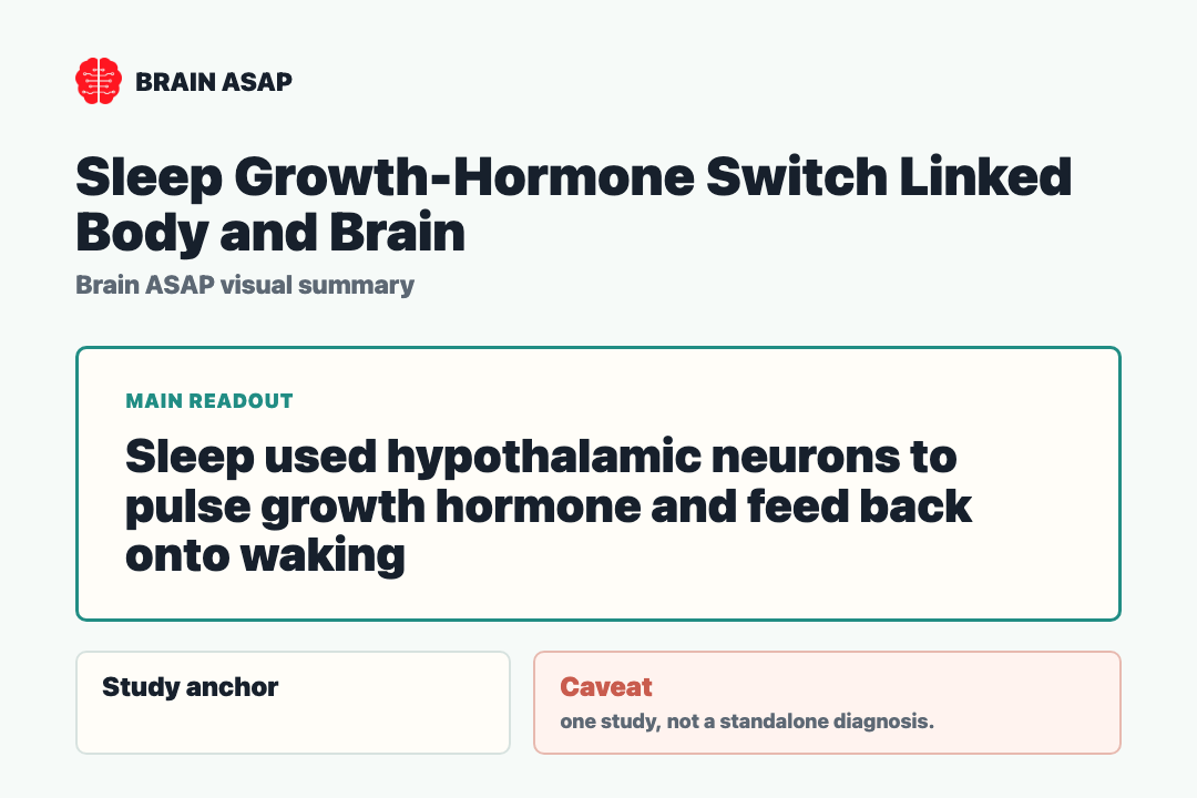
TL;DR: A 2025 mouse study in Cell mapped a sleep-growth hormone circuit, showing how hypothalamic GHRH and somatostatin neurons coordinate hormone pulses and then feed back onto wakefulness.
Key Findings
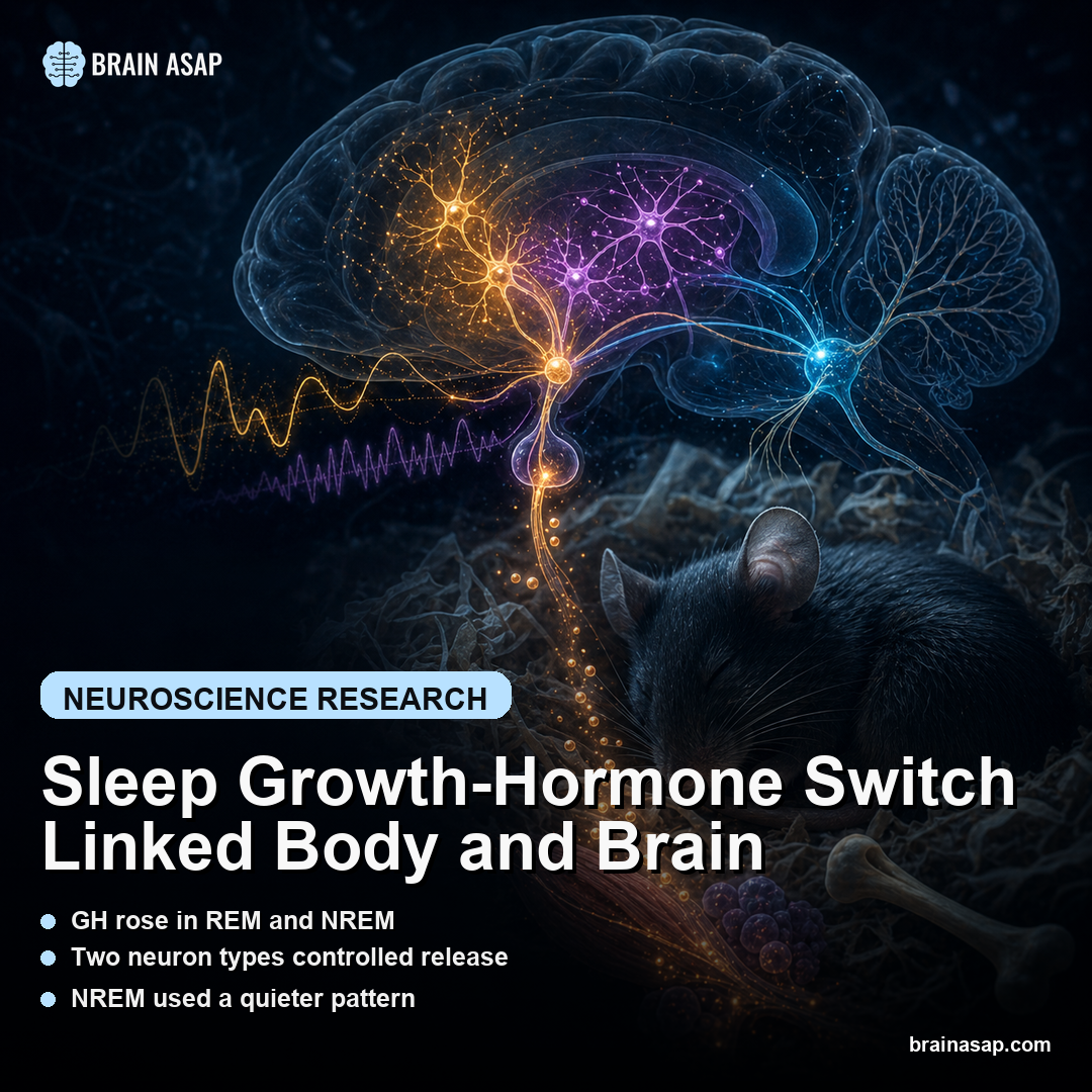
GH rose in REM and NREM: Growth hormone release was enhanced during both rapid eye movement and non-REM sleep.
Two neuron types controlled release: Hypothalamic GHRH neurons stimulated GH release, while somatostatin neurons suppressed it through distinct routes.
NREM used a quieter pattern: During NREM sleep, GHRH activity increased moderately while SST activity decreased.
REM produced strong surges: REM sleep was associated with strong surges of both GHRH and SST activity.
GH pushed back toward waking: Growth hormone enhanced locus coeruleus excitability and increased wakefulness, forming a negative feedback loop.
Source: Cell (2025) | Ding et al.
Sleep is not only a shutdown state. It is a hormonal control room, and growth hormone is one of its most consequential outputs for tissue growth, fat metabolism, and glucose regulation.
Growth Hormone Was the Sleep Output to Explain
Clinicians have known for decades that growth hormone release is tightly linked to sleep. What was missing was a circuit-level account of how the sleeping brain controls the pulse.
The study focused on hypothalamic neurons that either promote or suppress growth hormone release. The work is therefore less about sleep as a single state and more about sleep-stage-specific endocrine control.
The distinction is important for metabolism.
Growth hormone affects glucose handling, fat use, tissue growth, and repair.
If different sleep stages drive different hormone-control patterns, then disrupted sleep architecture can alter the body’s endocrine timing even when total sleep duration looks acceptable.
The study therefore connects two fields that are often discussed separately: sleep neurobiology and metabolic hormone regulation.
GHRH Promoted Release While Somatostatin Restrained It
Growth-hormone-releasing hormone neurons do what their name promises: they stimulate release. Somatostatin neurons restrain the system, but the paper separates at least two routes.
Arcuate SST neurons suppress nearby GHRH neurons, while periventricular SST neurons project to the median eminence. The finding is a circuit with both local inhibition and output-gate control.
The anatomy gives the circuit more nuance than a simple accelerator-brake model. Some somatostatin signaling appears to counteract GHRH neurons directly, while another branch controls hormone output closer to the endocrine interface.
This layered arrangement lets the brain tune hormone pulses rather than merely switch them on or off. It also gives researchers multiple nodes to test when sleep and metabolism drift out of sync.
REM and NREM Used Different Hypothalamic Patterns
During NREM sleep, GHRH activity rose moderately while SST activity fell. That sleep-stage arrangement makes intuitive sense for allowing hormone release.
REM sleep was stranger: both GHRH and SST activity surged. The circuit is not a simple on-off switch; it is a timed regulator that can produce hormone release while balancing feedback and restraint.
- NREM sleep: moderate GHRH activation paired with lower SST activity created a permissive hormone-release state.
- REM sleep: stronger surges in both GHRH and SST showed a more complex push-pull pattern.
- Hypothalamus: the control logic sat in specific hormone-regulating neuronal populations.
- Feedback: growth hormone then acted back on wake-promoting circuitry.
The REM result is informative because it warns against flattening sleep into one biological category. REM and NREM are not just different brain-wave states; in this study, they used different hypothalamic patterns to shape the same endocrine output.
Growth Hormone Fed Back Into the Locus Coeruleus
The most elegant piece is the loop back to the locus coeruleus, a wakefulness-related noradrenergic region. Growth hormone increased the excitability of locus coeruleus neurons and promoted waking.
That gives the system a brake. Sleep promotes hormone release, and hormone release then helps nudge the animal away from continued sleep.
This feedback loop makes biological sense.
Hormone release is informative, but the organism still has to move between sleep and wake states.
A growth-hormone signal that increases locus coeruleus excitability gives the system a way to limit the sleep period after endocrine needs are met.
It also connects a peripheral hormone to a central arousal circuit. The hormone is not only an output of sleep; it becomes part of the control loop that shapes what happens next.
Sleep Stages Carried Endocrine Timing Signals
Growth hormone touches sugar handling, fat metabolism, tissue repair, and muscle growth. Poor sleep can therefore affect the body through endocrine timing, not only through fatigue or appetite.
The translational promise is still early. But a circuit map gives researchers concrete nodes to test in sleep disorders that overlap with diabetes, obesity, Parkinson’s disease, or Alzheimer’s disease.
The mouse model caveat is important.
This mouse study does not prove that manipulating GHRH, somatostatin, or growth hormone feedback will treat human sleep or metabolic disease.
It provides a circuit logic that can guide the next experiments.
The result changes what sleep measurement should include.
Sleep is often measured by duration, fragmentation, or stages.
This study argues that endocrine timing should sit beside those measures when researchers ask how sleep changes the body.
For brain health, the locus coeruleus link is especially notable.
That region is involved in arousal, attention, stress physiology, and vulnerability in several neurodegenerative discussions.
A growth-hormone feedback signal into that system adds another bridge between metabolism and neural state.
The sleep physiology is not one-way. The brain shapes growth hormone release during sleep, and the hormone feeds back onto wake-promoting circuitry.
The study also gives sleep researchers a sharper target than “better sleep.” If a disorder fragments REM, alters NREM depth, or changes transitions into wakefulness, the endocrine consequence could differ. Stage timing may matter as much as the number of hours in bed.
Patients rarely present with sleep problems in isolation. Diabetes, obesity, stress, aging, and neurodegenerative risk often travel together, and a hypothalamic growth-hormone circuit gives those overlaps a plausible biological route.
The mouse data cannot settle the human question, but it can organize it. Future studies can ask whether altered growth-hormone timing tracks with sleep architecture, locus coeruleus-related arousal, and metabolic markers in people.
The finding is direct: sleep is an active endocrine program, not just a rest interval.
A informative human follow-up would measure sleep stages, growth-hormone pulses, arousal markers, and metabolic readouts in the same participants. That would move the field beyond treating sleep quality and hormone timing as separate outcomes.
The work also encourages a different reading of insomnia, sleep fragmentation, and circadian disruption.
The problem may not be only that the brain feels unrested.
The body may also miss or mistime endocrine signals that normally arrive during specific sleep states.
Growth hormone is not the whole explanation for sleep’s health effects. This circuit gives the field one concrete bridge between neural state and metabolic consequence.
The mouse result makes sleep stages look like active biological instructions. In this study, one instruction told the body when to release growth hormone, and the hormone then helped push the brain back toward wakefulness.
That feedback design also gives the system a built-in stopping rule. A hormone pulse can help complete a sleep-linked body program while also nudging arousal circuitry toward the next state.
The finding is a tidy example of why brain and body cannot be cleanly separated in sleep science. The sleeping brain controls the hormone, and the hormone talks back.
Citation: DOI: 10.1016/j.cell.2025.05.039. Ding et al.. Sleep-wake-dependent hypothalamic control of growth hormone release. Cell. 2025.
Study Design: Preclinical circuit study recording and manipulating sleep-wake-dependent hypothalamic neurons in mice.
Sample Size: Mouse experiments; exact animal counts vary by assay and should be checked at figure level.
Key Statistic: GH release was enhanced during REM and NREM sleep, with REM showing strong GHRH and SST surges and NREM showing moderately increased GHRH with decreased SST.
Caveat: Mouse circuit findings do not prove that manipulating the same pathway will improve human sleep or endocrine disease.