TL;DR: A 2026 medRxiv preprint used total-body tau PET in Alzheimer’s disease and found stronger brain-organ network synchrony, a pattern that tracked cognitive impairment through cortical tau burden and MRI markers of glymphatic dysfunction.
Key Findings
- Small imaging cohort: Researchers compared 28 Alzheimer’s disease patients with 23 matched negative controls using positron emission tomography (PET), MRI, and cognitive testing.
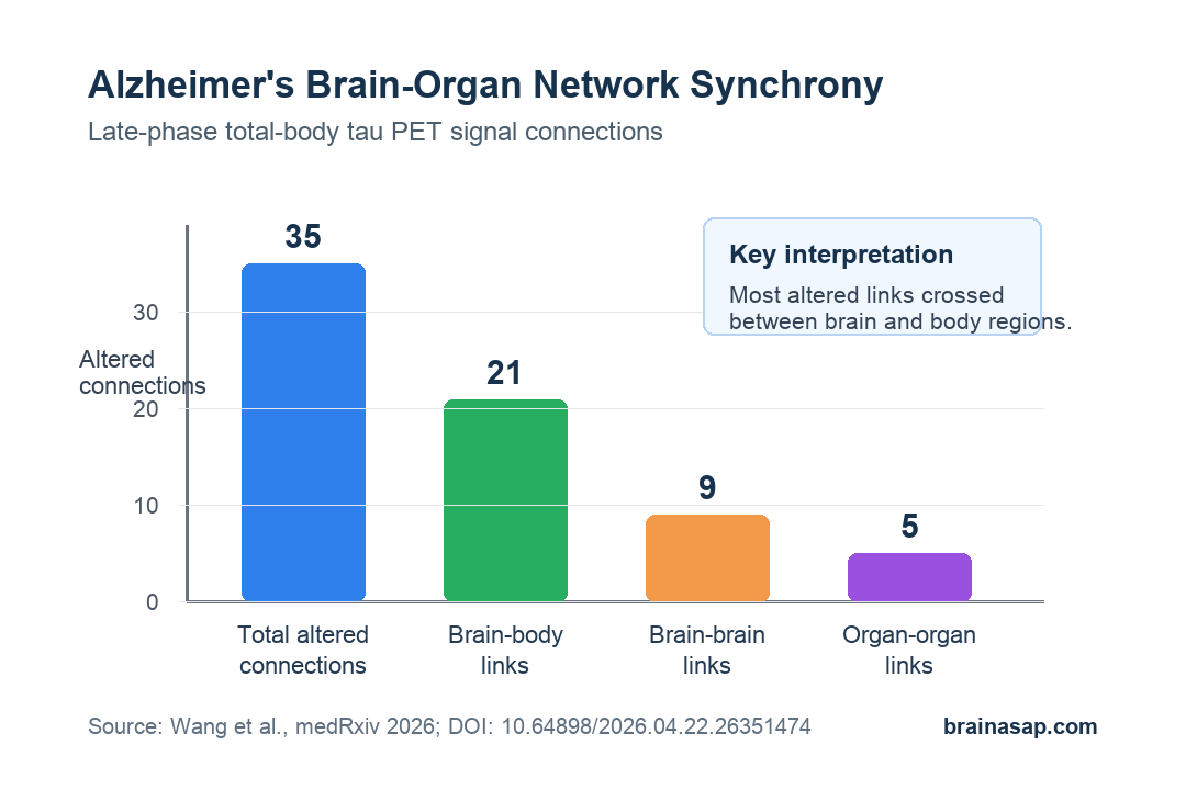
- Network pattern: Alzheimer’s disease was linked to 35 stronger brain-organ connections, including 21 brain-body links, after adjustment for age, sex, and education.
- Cognition pathway: The brain-organ connectivity score correlated with cortical tau burden (r = 0.52) and glymphatic MRI scores.
- MMSE link: MMSE, the Mini-Mental State Examination, was lower when the tau-burden score was higher.
- MoCA link: MoCA, the Montreal Cognitive Assessment, showed a similar negative association with the tau-burden score.
Source: medRxiv, 2026. This is a preprint and has not been peer reviewed.
Alzheimer’s disease is usually framed around the brain: amyloid plaques, tau tangles, memory circuits, and cortical atrophy.
This preprint asked a broader imaging question. If Alzheimer’s disease also changes systemic physiology, can whole-body PET detect abnormal timing relationships between the brain and organs?
Researchers used total-body 18F-tau PET, an imaging method that can follow tracer behavior across the brain and extracranial tissues during one scan.
They then built a brain-organ network (BON), meaning a map of how strongly low-frequency PET fluctuations moved together across brain regions and body organs.
Total-Body Tau PET Mapped Brain-Organ Network Synchrony
The study included 28 people with Alzheimer’s disease and 23 negative controls matched on age, sex, and education. The Alzheimer’s group had positive amyloid and tau PET findings, while the control group was negative for those pathologies.
The groups were clearly separated on cognitive testing:
- MMSE: The Mini-Mental State Examination averaged 14.9 in Alzheimer’s participants and 25.9 in controls.
- MoCA: The Montreal Cognitive Assessment averaged 9.4 in Alzheimer’s participants and 22.2 in controls.
For the network analysis, researchers extracted late-phase PET signal time courses from:
- Ten cortical regions: bilateral frontal, temporal, parietal, occipital, and insular cortices.
- Nine extracranial regions: kidneys, liver, lungs, colon, spleen, skeleton, heart, spinal cord, and skeletal muscles.
- Adjusted comparisons: group differences controlled for age, sex, and education.
The result went beyond higher cortical tau. Alzheimer’s disease also showed stronger synchronized PET fluctuations across the brain-organ network.
Researchers identified 35 stronger connections in the Alzheimer’s group: 9 brain-brain, 5 organ-organ, and 21 brain-body links.
Spinal Cord and Occipitotemporal Cortex Were Network Hubs
The altered network was not evenly spread across every region. The right occipital and temporal cortices stood out among brain nodes, while the spinal cord stood out among extracranial nodes.
Kidneys, liver, skeleton, and heart also appeared in the altered network.
The specific altered links help explain why the finding is more than a generic “whole body” claim:
- Brain-brain links: stronger differences appeared around bilateral occipital regions and right frontal-temporal connections.
- Organ-organ links: the spinal cord showed altered coupling with lungs, skeletal muscle, and kidneys.
- Brain-body links: right occipital cortex connected differently with liver, right temporal cortex connected differently with skeletal muscle and heart, and spinal cord coupling changed with left parietal cortex.
Static tau measurements showed higher tau uptake in major cortical lobes, but not in extracranial organs.
The body pattern in this paper was mainly a dynamic synchrony finding, not evidence that tau deposits were higher throughout the body.
Tau Burden Linked Network Synchrony to MMSE and MoCA Scores
Researchers summarized cortical tau burden into a principal-component score called SUVR-PC. SUVR means standardized uptake value ratio, a PET measure that compares tracer uptake in a target region with a reference region.
They also summarized the altered brain-organ connections into a BON-PC score.
In the Alzheimer’s group, the two scores were moderately related. Higher BON-PC tracked higher SUVR-PC with a partial correlation of r = 0.52 after adjustment for age, sex, and education.
Higher tau burden also tracked worse cognition, with correlations of r = -0.56 for MMSE and r = -0.46 for MoCA.
The mediation models were exploratory, but they gave the network result a clearer structure:
- Network to tau: higher brain-organ connectivity predicted higher cortical tau burden.
- Tau to cognition: higher tau burden predicted lower MMSE and MoCA scores.
- Direct network path: after tau was included, the direct BON-to-cognition path was no longer statistically significant.
This statistical model does not prove a causal chain. It means the cross-sectional data fit a model where abnormal brain-organ synchrony shares variance with cognitive impairment through cortical tau burden.
Glymphatic MRI Markers Added a Second Cognition Pathway
The study also tested MRI-derived markers related to the glymphatic system, the brain’s fluid-clearance pathway for cerebrospinal and interstitial fluid exchange.
Researchers combined 11 MRI indices into glymphatic summary scores:
- Perivascular-space measures: volume fractions in whole brain, white matter, and basal ganglia.
- Free-water measures: diffusion-derived estimates across brain tissue compartments.
- Fluid-dynamics measures: ALPS diffusion indices and BOLD-CSF coupling.
The brain-organ network score correlated with two glymphatic summary scores. The stronger association was with GLY-PC2 at r = 0.61, while GLY-PC1 showed r = 0.40.
GLY-PC1 then appeared in mediation models linking stronger brain-organ synchrony with lower cognition scores.
In a parallel model, cortical tau burden and glymphatic dysfunction each carried independent statistical links between BON-PC and cognition.
For MMSE, both pathways were significant and the direct BON-PC path became non-significant. For MoCA, the direct path was attenuated but still reached significance.
What This Alzheimer’s PET Preprint Can and Cannot Show
The strongest contribution is methodological. The paper shows how total-body PET can be used to study Alzheimer’s disease as a network of brain and body physiology, not only as a static map of cortical tau uptake.
The limits are equally important:
- Preprint status: the analysis has not been peer reviewed.
- Small sample: the core cohort was 51 people, with 28 Alzheimer’s patients.
- Cross-sectional design: the mediation results are statistical pathways, not proof that brain-organ synchrony causes tau accumulation or cognitive decline.
- Mechanism still open: the PET synchrony could reflect vascular, autonomic, tracer-exchange, inflammatory, or fluid-clearance processes that need prospective testing.
If the finding replicates, Alzheimer’s imaging may need to pay closer attention to brain-body timing patterns, not just whether a tracer is high in one tissue.
For now, the finding maps a hypothesis about systemic physiology in Alzheimer’s disease.
Citation: DOI: 10.64898/2026.04.22.26351474. Wang et al. Brain-Organ Hypersynchrony and Cognitive Decline in Alzheimer’s Disease: Potential Links with Tauopathy and Glymphatic Dysfunction. medRxiv. 2026.
Study Design: Single-center retrospective total-body PET and MRI analysis comparing Alzheimer’s disease patients with negative controls.
Sample Size: 28 Alzheimer’s disease patients and 23 amyloid/tau-negative controls.
Key Statistic: Alzheimer’s disease was associated with 35 stronger brain-organ network connections, including 21 brain-body links.
Caveat: This was a small cross-sectional preprint, so mediation paths should be treated as statistical associations rather than causal evidence.