TL;DR: A 2026 functional MRI (fMRI) study in Psychological Medicine found that schizophrenia, bipolar disorder, and major depressive disorder shared some thalamus connectivity disruption, but each diagnosis showed a different subcortical network pattern across the thalamus, striatum, hippocampus, and amygdala.
Key Findings
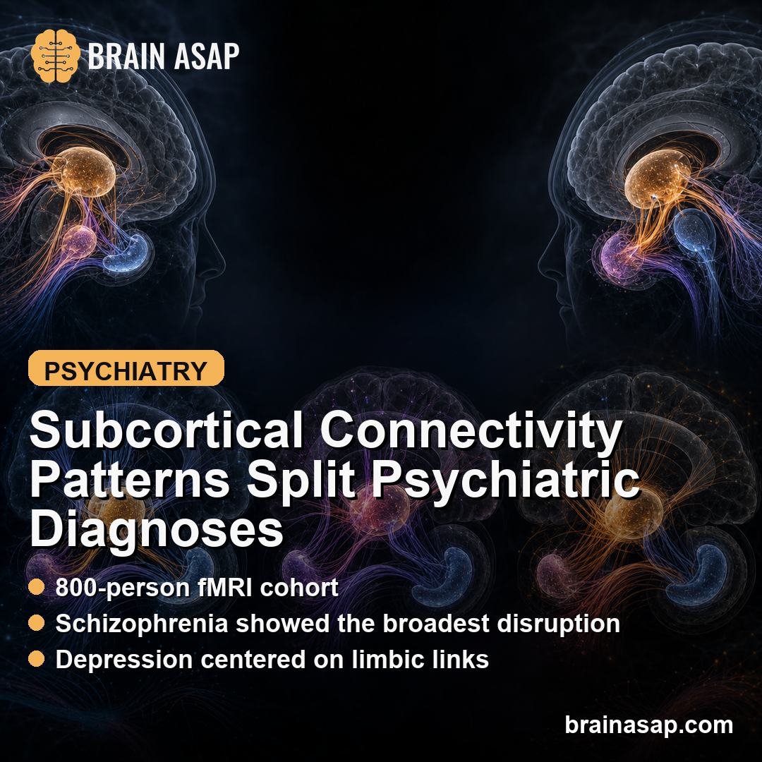
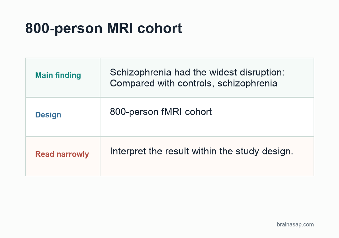
- 800-person MRI cohort: Researchers analyzed resting-state fMRI from 200 people with schizophrenia, 200 with bipolar disorder, 200 with major depressive disorder, and 200 healthy controls at a single Taiwan site.
- 54 subcortical regions: The analysis divided the thalamus, striatum, hippocampus, and amygdala into 54 regions and tested 1,431 pairwise functional-connectivity comparisons for each diagnostic contrast.
- Schizophrenia had the widest disruption: Compared with controls, schizophrenia showed abnormal connectivity in 17.3% of tested subcortical connections, including 54 reduced intra-thalamic links and 104 increased intra-striatal links.
- Bipolar disorder showed a different striatal direction: Bipolar disorder also had reduced intra-thalamic connectivity, but its striatal pattern leaned toward reduced rather than increased connectivity.
- Depression was more focal: Major depressive disorder showed abnormal connectivity in 1.6% of tested connections, with the clearest reductions inside the hippocampus-amygdala complex and between striatal and limbic regions.
Source: Psychological Medicine (2026) | Chang et al.
Subcortical Networks Were Compared Across Three Diagnoses
Subcortical brain regions sit below the cerebral cortex and help route sensory, emotional, motivational, and cognitive signals. This study focused on four connected systems: the thalamus, striatum, hippocampus, and amygdala.
The question was not whether one diagnosis had a single abnormal brain region. Researchers asked whether the internal wiring of these subcortical systems differed across schizophrenia, bipolar disorder, and major depressive disorder.
These diagnoses can overlap clinically. Psychosis, mood symptoms, cognitive problems, and motivational changes do not map neatly onto one brain circuit.
A high-resolution connectivity approach can show where the same broad symptom territory may still come from different network patterns.
- Thalamus: A relay and gating hub involved in sensory, cognitive, and cortical communication.
- Striatum: A basal-ganglia system tied to motivation, reward, movement, habit learning, and dopaminergic signaling.
- Hippocampus-amygdala complex: A limbic system involved in memory, threat processing, emotional salience, and mood regulation.
Researchers Tested 1,431 Subcortical Connectivity Pairs
The dataset included 800 participants, evenly split into four groups of 200: schizophrenia, bipolar disorder, major depressive disorder, and healthy controls. All participants came from one site, which reduces scanner and protocol variability compared with multi-site pooling.
Researchers used resting-state functional MRI, a method that estimates how strongly brain regions fluctuate together when a person is not performing a task. In plain terms, functional connectivity means that two regions show synchronized activity patterns over time.
The analysis used a detailed atlas that separated subcortical structures into 54 regions: 8 thalamus regions, 12 striatum regions, 5 hippocampus regions, and 2 amygdala regions per hemisphere. Pairwise connectivity was then tested among these regions.
- Within-structure links: Connections inside the thalamus, inside the striatum, and inside the hippocampus-amygdala complex.
- Between-structure links: Connections between basal ganglia, thalamus, and hippocampus-amygdala regions.
- Clinical severity checks: Exploratory analyses tested whether within-structure connectivity tracked symptom severity scores.
Schizophrenia Showed Thalamic Loss and Striatal Overconnectivity
The schizophrenia group had the broadest subcortical disruption. Compared with healthy controls, abnormal connectivity appeared in 17.3% of tested connections, far more than in the bipolar or depression groups.
The clearest within-structure pattern was a split: 54 intra-thalamic connections were reduced, while 104 intra-striatal connections were increased. The thalamus looked less internally coordinated, while the striatum looked more synchronized than expected.
Between structures, schizophrenia also showed widespread increases among thalamus, striatum, and limbic regions. The broader subcortical communication shift was not isolated to one region.
Clinical correlations added another layer. In schizophrenia, higher intra-striatal connectivity correlated with greater psychotic symptom severity, although the correlations were small.
Researchers also noted that antipsychotic treatment could contribute to some striatal and between-region findings, so the schizophrenia pattern should not be read as a medication-free disease signature.
Bipolar Disorder Shared Thalamic Disruption but Reversed the Striatal Pattern
Bipolar disorder also showed intra-thalamic hypoconnectivity, meaning reduced functional connectivity among thalamus regions. In this study, 29 intra-thalamic links were reduced relative to healthy controls.
The striatum moved in the opposite direction from schizophrenia. Bipolar disorder showed 11 reduced intra-striatal connections, not the large striatal increase seen in schizophrenia.
The opposite striatal direction is one of the study’s clearest diagnostic contrasts.
Between structures, bipolar disorder showed reduced connectivity between thalamus and striatum, and between striatum and limbic structures. One notable finding was reduced amygdala-putamen connectivity, with a peak t value of -4.17 and p < 0.0001.
- Shared feature: Both schizophrenia and bipolar disorder showed reduced thalamus internal connectivity.
- Opposite striatal direction: Schizophrenia showed striatal hyperconnectivity, while bipolar disorder showed striatal hypoconnectivity.
- Medication caveat: Between-region subcortical links were more sensitive to antipsychotic medication status than the core within-structure findings.
Depression Centered More on Limbic and Striatum-Limbic Links
Major depressive disorder had a more limited connectivity footprint. Abnormal connectivity appeared in 1.6% of tested connections, compared with 17.3% for schizophrenia and 7.1% for bipolar disorder.
The strongest depression pattern was reduced connectivity within the hippocampus-amygdala complex. Researchers reported reduced links between left medial and lateral amygdala, between left medial amygdala and right lateral amygdala, and between left and right hippocampus.
Depression also showed reduced connectivity between striatal and limbic regions, especially amygdala-putamen connectivity, where the peak t value was -4.4 with p < 0.00001. That fits a clinically plausible circuit: reward and motivation systems interacting with emotional-salience regions.
Only one intra-thalamic connection was significantly reduced in the depression group. The study therefore supports a cautious reading: depression may share some thalamic disruption, but its main subcortical signature here was more limbic and striatum-limbic than globally thalamic.
The Findings Are Network Clues, Not Diagnostic Brain Scans
This study should not be read as a clinical MRI test for any one patient. The analysis compared groups, and the participants had demographic and treatment differences.
The major depressive disorder group was older on average, more female, and had shorter illness duration than the schizophrenia and bipolar groups.
Medication is another real limitation. Antipsychotics, antidepressants, and mood stabilizers were common across patient groups.
The sensitivity analysis suggested that within-subcortical abnormalities were more stable than between-structure abnormalities, but treatment effects cannot be fully removed from an observational imaging study.
Still, the paper gives a useful map of how serious psychiatric disorders can overlap and diverge below the cortex. A simplified reading is:
- Schizophrenia: Broad subcortical dysconnectivity, with thalamic reduction and strong striatal increase.
- Bipolar disorder: Thalamic reduction plus reduced striatal and striatum-limbic connectivity.
- Major depressive disorder: More focal limbic and striatum-limbic reductions, with only mild thalamic involvement.
These findings do not support diagnosis by scan. They give researchers a concrete framework for studying shared symptoms, treatment response, and why psychiatric categories can look similar at the surface while differing in subcortical network biology.
Citation: DOI: 10.1017/S0033291726103377. Chang et al. Distinct and common subcortical functional connectivity revealed across three major psychiatric disorders. Psychological Medicine. 2026;56:e109.
Study Design: Single-site resting-state functional MRI group comparison across three psychiatric diagnoses and healthy controls.
Sample Size: 800 participants: 200 with schizophrenia, 200 with bipolar disorder, 200 with major depressive disorder, and 200 healthy controls.
Key Statistic: Abnormal subcortical connectivity appeared in 17.3% of tested connections for schizophrenia, 7.1% for bipolar disorder, and 1.6% for major depressive disorder.
Caveat: The study was observational, group-level, and medication exposure differed across diagnostic groups.