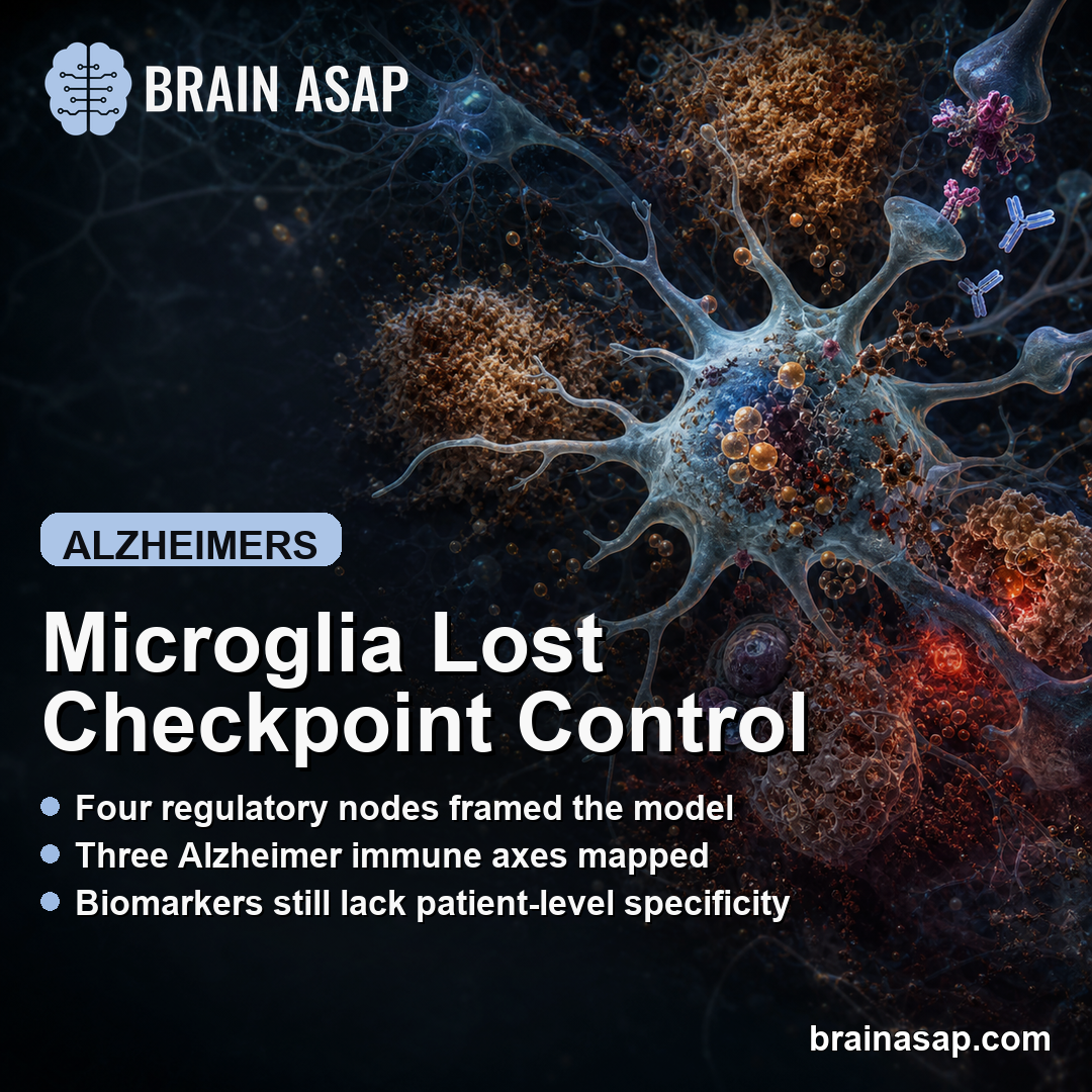
TL;DR: A 2026 review in Journal of Neuroinflammation proposed that Alzheimer microglial dysfunction can be understood as checkpoint collapse, where failed regulatory controls feed three interacting lipid, iron/ferroptosis, and inflammation/complement axes.
Key Findings
- Review framework, not trial result: Alzheimer microglial evidence through April 2026 was synthesized rather than used to test a new drug or diagnostic rule.
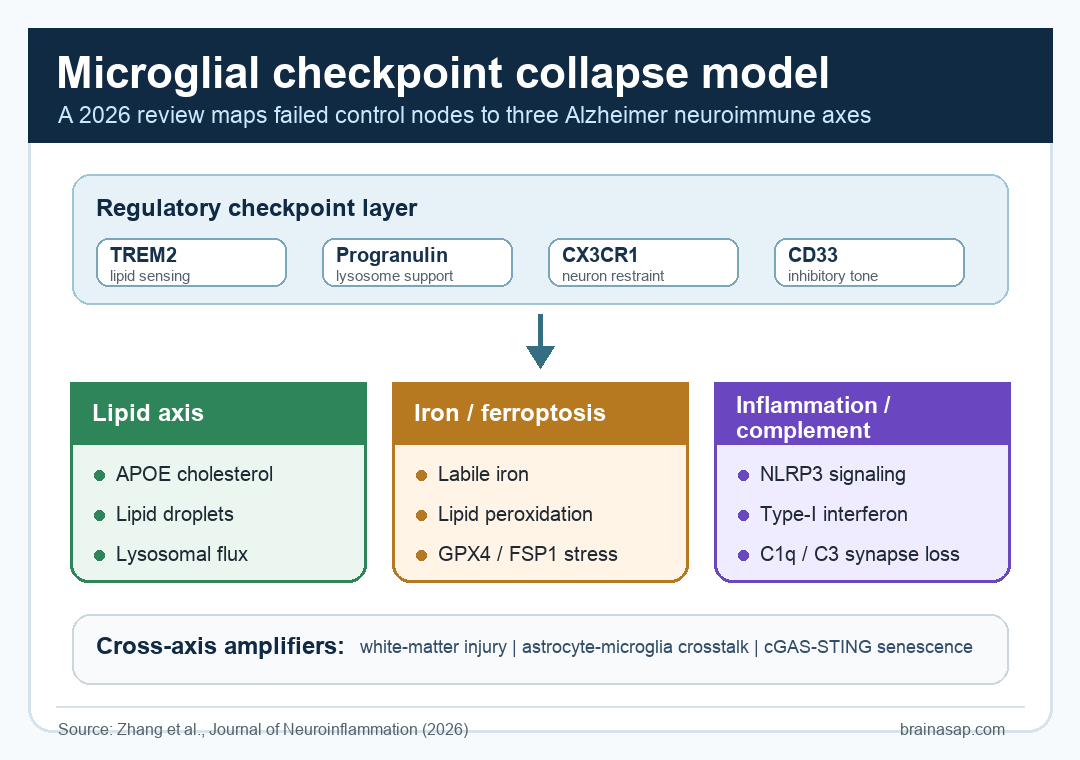
- Four checkpoint nodes: The framework centers on TREM2 lipid sensing, progranulin-linked lysosomal function, CX3CR1 neuron-microglia restraint, and CD33/Siglec-3 inhibitory tone.
- Three downstream axes: Failed regulation was mapped onto lipid handling, iron/ferroptosis stress, and inflammation/complement activity.
- Cross-axis amplifiers: White-matter injury, astrocyte-microglia crosstalk, and cGAS-STING-linked senescence were treated as amplifiers rather than separate isolated pathways.
- Clinical caution: The authors emphasized that current glial, iron, inflammatory, and imaging biomarkers are not specific enough to assign individual patients to one axis in routine care.
Source: Journal of Neuroinflammation (2026) | Zhang et al.
Anti-Amyloid Therapy Left Microglial Biology Unsettled
Anti-amyloid antibodies have made one point clearer: amyloid beta is biologically relevant in Alzheimer disease.
The review starts from the harder follow-up problem. Removing fibrillar amyloid does not automatically settle the glial, lipid, vascular, and inflammatory processes that continue around plaques, tau pathology, and damaged tissue.
That is where microglia become central. These immune cells help clear debris, remodel synapses, process lipid-rich material, and regulate local inflammatory thresholds.
In Alzheimer disease, they sit near plaques, dystrophic neurites, oxidized lipids, injured myelin, and inflammatory signals.
Generic “activation” language is too blunt for this biology. Some microglial responses may start as protective attempts to contain damage.
The problem is what happens when the regulatory controls that keep those responses useful begin to fail.
Four Regulatory Nodes Form the Checkpoint Layer
Checkpoint collapse is the review’s name for progressive loss of microglial transition control. The idea is not that one switch breaks.
It is that several control nodes lose their ability to keep microglia inside a recoverable, tissue-protective range.
The framework highlights four main regulatory systems:
- TREM2: Supports lipid and apolipoprotein sensing, phagocytic competence, and metabolic adaptation under chronic plaque-related load.
- Progranulin: Helps maintain lysosomal acidification and degradative capacity when microglia are handling aggregate and lipid-rich cargo.
- CX3CR1: Provides neuron-derived restraint through fractalkine signaling, limiting excessive microglial reactivity and synaptic engulfment.
- CD33/Siglec-3: Contributes inhibitory tone that helps set innate immune activation thresholds.
Those nodes do different jobs, but they point to the same concept. Alzheimer microglial dysfunction is not simply “too much inflammation.” It may reflect weaker control over lipid cargo, lysosomal clearance, neuron-microglia signaling, and inflammatory triggering at the same time.
Three Effector Axes Organize the Downstream Damage
Once the regulatory layer weakens, downstream pathology can be organized into three interacting axes. The axes are separated because they point to different experiments, biomarkers, and drug strategies, but they remain connected biology rather than clean compartments.
- Lipid axis: APOE-biased cholesterol trafficking, impaired lysosomal flux, and lipid-droplet-accumulating microglia can leave cells less able to recycle plaque- and myelin-associated cargo.
- Iron/ferroptosis axis: Labile iron, peroxidized phospholipids, and insufficient GPX4/FSP1 defenses can push microglia toward oxidative injury programs that can harm nearby neurons.
- Inflammation/complement axis: NLRP3 activation, type-I interferon signaling, and C1q/C3-dependent synaptic engulfment can link immune activation to tau pathology and synapse loss.
That structure helps explain why lipid droplets, iron retention, complement activation, white-matter changes, and astrocyte reactivity often appear together in Alzheimer tissue. Lipid-handling failure can worsen lysosomal congestion and ferritin turnover.
Iron-driven lipid peroxidation can prime inflammasome pathways. Complement and inflammatory output can then feed back into lipid handling through astrocyte-microglia crosstalk.
Biomarkers Still Cannot Assign a Patient to One Axis
The framework is biomarker-facing, but it is deliberately cautious. The authors describe possible readouts for each axis while warning that none of them is specific enough to classify an individual patient into a discrete microglial subtype in routine practice.
Examples of axis-linked readouts include:
- Lipid vulnerability: APOE genotype plus plasma or cerebrospinal fluid (CSF) lipidomics, including ceramide, sphingomyelin, and neutral-lipid signatures.
- Iron/ferroptosis biology: QSM-MRI, CSF ferritin, and lipid-peroxidation measures that may show regional iron and oxidative membrane stress.
- Inflammation/complement activity: C1q, C3, inflammasome-linked measures, and microglial positron emission tomography (PET) tracer tools such as TSPO or newer CSF1R/TREM2-targeted tools.
- Glial amplification: sTREM2, progranulin-related measures, GFAP, and YKL-40, interpreted alongside amyloid, tau, vascular, and disease-stage context.
The important word is context. A higher GFAP or YKL-40 signal may indicate glial activation, but it does not tell a clinician by itself whether lipid handling, complement pruning, ferroptosis, or astrocyte response is the rate-limiting problem.
The review’s practical message is that biomarker panels should narrow hypotheses, not pretend to deliver one-axis certainty.
Treatment Ideas Need Stage and Biomarker Anchors
The review also uses the framework to discipline therapeutic thinking.
A combination should not mean stacking every plausible anti-inflammatory, anti-amyloid, antioxidant, and complement drug together. It should mean matching a stage, an axis, and a pharmacodynamic readout.
The stage logic is straightforward:
- Early disease: Regulatory-node preservation and selective inflammasome restraint may matter most if microglia still have recoverable control capacity.
- Intermediate disease: Lipid-handling restoration and ferroptosis control may become more relevant as chronic plaque, tau, and myelin stress accumulate.
- Later disease: Complement moderation, senescence-directed approaches, and white-matter or astrocyte-microglia context may become more central to target selection.
That is still a research framework, not a treatment manual. Some proposed strategies, such as TREM2 agonism, NLRP3 inhibition, complement targeting, ferroptosis modulation, or senescence-directed therapy, remain dependent on stage selection and proof of central target engagement.
The useful shift is conceptual precision. Instead of treating “neuroinflammation” as one broad target, the practical task becomes identifying which checkpoint failed, which axis is carrying the pathology, and which biomarker should move if the intervention is actually doing what it was designed to do.
Citation: DOI: 10.1186/s12974-026-03924-x. Zhang et al. Microglial checkpoint collapse in Alzheimer’s disease: a tri-axial framework for biomarker-informed neuroimmune therapy. Journal of Neuroinflammation. 2026.
Study Design: Narrative mechanistic review and translational framework synthesis, not a systematic review or clinical trial.
Sample/Model: Human Alzheimer tissue studies, single-cell and spatial profiling, genetic evidence, animal and cell models, biomarker studies, and clinical-intervention reports.
Key Statistic: The review mapped microglial dysfunction onto checkpoint collapse plus three coupled effector axes: lipid handling, iron/ferroptosis, and inflammation/complement.
Caveat: The framework is hypothesis-generating; current biomarkers cannot reliably assign individual patients to discrete microglial axes in clinical practice.