TL;DR: A 2026 systematic review and meta-analysis in Journal of Alzheimer’s Disease found that choroid plexus volume, an MRI-visible structure involved in cerebrospinal-fluid regulation and immune signaling, was larger in Alzheimer’s disease than in healthy controls.
Key Findings
- Sixteen studies: The review included 16 MRI-based studies covering 2,004 Alzheimer’s disease patients and 883 healthy controls.
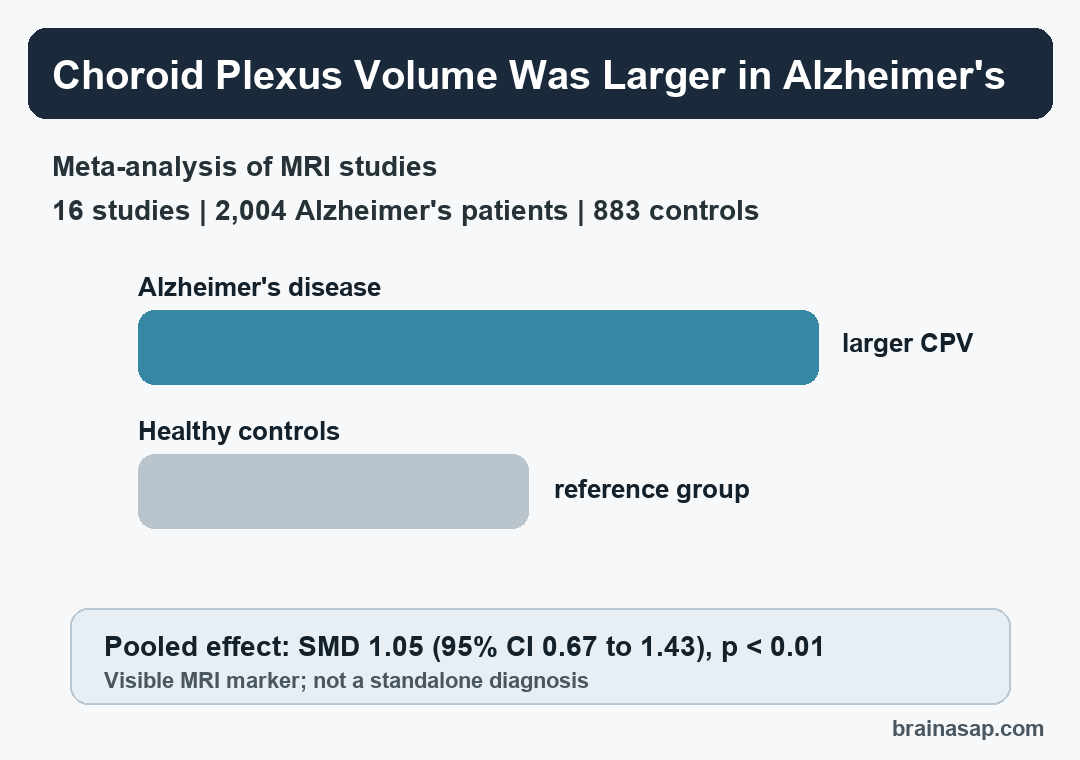
- Larger choroid plexus: Pooled results showed higher choroid plexus volume in Alzheimer’s disease, with SMD 1.05 and a 95% confidence interval from 0.67 to 1.43.
- Cognitive links: Narrative findings connected larger choroid plexus volume with lower Mini-Mental State Examination scores and worse global cognition.
- Pathology context: Reported associations also included hippocampal and cortical atrophy, ventricular expansion, amyloid and tau deposition, and impaired glymphatic clearance.
- Important caveat: Choroid plexus enlargement is not Alzheimer’s-specific; the review frames it as a marker of clearance, inflammation, and barrier dysfunction rather than a standalone diagnosis.
Source: Journal of Alzheimer’s Disease (2026) | Mahmoudi et al.
The choroid plexus sits inside the brain’s ventricles and helps produce cerebrospinal fluid. It also forms part of the blood-cerebrospinal-fluid barrier, a boundary that affects protein clearance and immune traffic between the body and central nervous system.
The structure is relevant to Alzheimer’s disease for a practical reason. Alzheimer’s pathology is usually described through amyloid beta, tau, brain atrophy, and cognitive decline, but the brain also has to clear proteins, regulate fluid movement, and manage inflammation.
This review asked whether the choroid plexus shows a reproducible MRI finding in Alzheimer’s disease. The pooled comparison found larger volume in Alzheimer’s disease, with an important limitation: the measurement is better read as a clearance-and-inflammation clue than as a disease-specific fingerprint.
Alzheimer’s Disease Showed Larger Choroid Plexus Volume on MRI
Researchers searched PubMed, Embase, Scopus, and Web of Science through March 2025. Eligible studies had to use MRI and compare choroid plexus volume between Alzheimer’s disease patients and healthy controls, or evaluate how that volume related to clinical and biological features.
The pooled comparison included 16 studies, 2,004 Alzheimer’s disease patients, and 883 healthy controls. Because studies measured volume in different ways, the analysis used standardized mean difference, a method that lets results be compared across different units and normalization approaches.
The main result was large and statistically clear. Choroid plexus volume was higher in Alzheimer’s disease, with SMD 1.05, 95% CI 0.67 to 1.43, and p < 0.01.
The result also survived a leave-one-out sensitivity analysis. In plain terms, removing one study at a time did not erase the overall finding.
Larger Choroid Plexus Volume Tracked Cognition and Brain Pathology
The review could not pool every secondary association because the available datasets were too varied. Instead, the analysis summarized reported relationships across studies.
Those findings make the choroid plexus result more clinically relevant, while also keeping it less definitive than the main volume comparison.
Across the narrative synthesis, larger choroid plexus volume was linked with several Alzheimer’s-relevant features:
- Worse cognition: Studies reported lower Mini-Mental State Examination scores, worse global cognition, and poorer verbal learning performance.
- More structural injury: Larger volume appeared alongside reduced hippocampal and cortical volumes, lateral ventricular expansion, and periventricular white-matter hyperintensities.
- More Alzheimer’s pathology: Some studies connected choroid plexus enlargement with higher amyloid and tau deposition.
- Clearance and inflammation markers: Reported links included lower glymphatic-function measures and higher peripheral inflammatory markers.
The review also noted that choroid plexus changes were seen in mild cognitive impairment. Mild cognitive impairment can sit between normal aging and dementia, though not everyone with mild cognitive impairment progresses to Alzheimer’s dementia.
Choroid Plexus Volume Is Not a Standalone Alzheimer’s Test
The strongest practical reading is not that MRI choroid plexus volume can diagnose Alzheimer’s disease by itself. The review is more careful.
Choroid plexus volume is a macrostructural MRI measure, not a direct measurement of cerebrospinal-fluid flow, barrier function, or immune-cell entry.
Several interpretation problems remain:
- High heterogeneity: The main pooled result had I2 = 88%, meaning study results varied substantially even though the overall direction was consistent.
- Different MRI methods: Studies differed in scanner strength, acquisition protocols, segmentation methods, and volume normalization.
- Cross-sectional evidence: Most data could show association, not whether choroid plexus enlargement drives disease, compensates for disease, or follows other pathology.
- Partial-volume risk: Ventricular enlargement can complicate choroid plexus measurement, especially in neurodegenerative disease.
The review also points out that choroid plexus enlargement has been reported in other conditions, including multiple sclerosis and schizophrenia. That weakens any disease-specific claim but supports a broader interpretation: the choroid plexus may be responding to inflammation, altered fluid dynamics, and barrier stress across brain disorders.
The Clearance Biology Is the Useful Part
The choroid plexus connects several Alzheimer’s disease mechanisms that are often discussed separately. It contributes to cerebrospinal-fluid production, blood-cerebrospinal-fluid exchange, immune surveillance, and the handling of proteins that can accumulate in the brain.
For Alzheimer’s research, the practical question is not whether the choroid plexus replaces amyloid or tau as a biomarker. It does not.
A more realistic question is whether routine MRI can capture one part of the disease environment that standard plaque-and-tangle framing misses.
That framing suggests three possible uses for future research:
- Disease staging: Choroid plexus volume might help describe where a person sits along a clearance-inflammation continuum.
- Longitudinal monitoring: Repeated MRI could test whether choroid plexus changes track progression or treatment response.
- Biomarker combinations: Volume measures may be more useful when interpreted alongside amyloid, tau, cognition, atrophy, and inflammatory markers.
Those uses still need stronger evidence. The review calls for standardized segmentation, longitudinal studies, and clearer separation between Alzheimer’s-specific pathology and general aging or neuroinflammatory effects.
For now, the result makes the choroid plexus a clearer target for Alzheimer’s MRI research. It does not provide a diagnosis on its own, but it gives researchers a visible structure to study when asking how fluid clearance and immune regulation fit into Alzheimer’s disease.
Citation: DOI: 10.1177/13872877261447404. Mahmoudi et al. Choroid plexus volume in Alzheimer’s disease: A systematic review and meta-analysis. Journal of Alzheimer’s Disease. 2026;112(1):53-64.
Study Design: Systematic review and random-effects meta-analysis of MRI-based choroid plexus volume studies.
Sample Size: Sixteen studies, including 2,004 Alzheimer’s disease patients and 883 healthy controls.
Key Statistic: Alzheimer’s disease patients had larger choroid plexus volume than healthy controls, with SMD 1.05 and 95% CI 0.67 to 1.43.
Caveat: The evidence was heterogeneous and mostly cross-sectional, so choroid plexus volume should not be treated as an Alzheimer’s-specific diagnostic marker.