TL;DR: A 2026 Communications Biology study associated maternal SARS-CoV-2 infection during pregnancy with more positive toddler autism-risk screens and newborn immune-protein patterns tied to nicotinamide metabolism, microglial activation, and neutrophil activity.
Key Findings
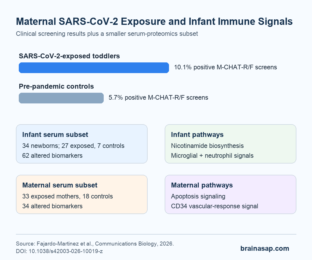
- Positive M-CHAT-R/F screens, a toddler autism-risk screening result, occurred in 10.1% of 218 SARS-CoV-2-exposed children versus 5.7% of 527 pre-pandemic controls.
- Earlier developmental-delay analysis in the same research program reported 11.6% delay in exposed children versus 1.6% in controls when Bayley-III and Ages and Stages Questionnaire results were included.
- Newborn serum profiling covered 34 infants, including 27 exposed newborns and 7 control newborns.
- Exposed infants with positive screens showed 62 altered serum biomarkers, with pathways involving nicotinamide biosynthesis, microglial activation, and neutrophil extravasation.
- Maternal serum profiling found 34 altered biomarkers in SARS-CoV-2-affected pregnancies, including apoptosis-related proteins such as CASP2, MAP3K5, FHIT, BIRC2, and BAX.
Maternal infection can affect brain development even when the fetus is not directly infected. Immune activation during pregnancy can change inflammatory messages reaching the placenta, fetal tissues, and developing nervous system.
This study followed that question through a COVID-19 pregnancy cohort. Researchers compared children exposed to laboratory-confirmed maternal SARS-CoV-2 infection during pregnancy with children born before the pandemic, then looked at immune proteins in a smaller subset of mother-infant blood samples.
This is not a diagnosis claim. The study connects prenatal infection history, toddler screening outcomes, and peripheral immune profiles.
SARS-CoV-2-Exposed Children Had More Positive Autism-Risk Screens
The clinical screening outcome used here was the Modified Checklist for Autism in Toddlers-Revised with Follow-Up, usually shortened to M-CHAT-R/F. It is a validated screening tool for autism risk in toddlers, not a final autism diagnosis.
Children were screened between 18 and 32 months. A positive screen led to referral for specialized diagnostic early-intervention services.
The main comparison was straightforward:
- SARS-CoV-2-exposed group: 22 of 218 children had a positive M-CHAT-R/F screen, or 10.1%.
- Pre-pandemic control group: 30 of 527 children had a positive M-CHAT-R/F screen, or 5.7%.
- Statistical result: the exposed group was nearly twice as likely to screen positive, with p = 0.03.
- Birth infection detail: none of the exposed infants tested positive for SARS-CoV-2 at birth.
That birth detail separates newborn infection from maternal immune exposure. The study focused on the pregnancy immune environment and the developmental follow-up of exposed children.
Vaccination and Sex Were Associated With Screen Results
Researchers also tested whether common pregnancy or infant factors explained the positive M-CHAT-R/F screens in the exposed group.
In adjusted models, male infant sex was associated with a higher likelihood of a failed M-CHAT-R/F result. Maternal vaccination before delivery was associated with a lower likelihood of a failed screen.
- Male infant sex: adjusted odds ratio 1.12, 95% CI 1.03 to 1.22, p = 0.01.
- Maternal vaccination before delivery: adjusted odds ratio 0.89, 95% CI 0.82 to 0.98, p = 0.02.
- Trimester of infection: first, second, or third trimester timing was not significantly associated with positive screens.
- Maternal comorbidities: hypertension, diabetes, obesity, and mental-health comorbidity did not explain the exposed-group screen pattern.
The vaccination result should be read carefully. It supports an association with lower screen risk in this cohort, not proof that vaccination directly changed neurodevelopmental outcomes.
Newborn Immune Proteins Pointed to Brain-Relevant Pathways
The mechanistic part of the paper used serum proteomic profiling, which measures many circulating proteins at once. The infant subset was small: 34 newborns, including 27 SARS-CoV-2-exposed infants and 7 controls.
Among exposed infants who later had positive M-CHAT-R/F screens, researchers found 62 altered serum biomarkers compared with healthy controls.
The enriched pathways were notable because several connect plausibly to fetal or early-life brain biology:
- Nicotinamide biosynthesis: proteins such as NMNAT1 and NADK relate to NAD-linked cellular energy and redox balance.
- Microglial activation: proteins including MMP8, ITGAM, AZU1, and AIF1 pointed toward immune-cell programs relevant to neuroinflammation.
- Neutrophil extravasation: SERPINB1, PRTN3, LBR, and NCF2 suggested altered innate immune trafficking and activation.
- Chemotactic responses: some cell-migration response pathways were downregulated, showing that the immune pattern was not simply global activation.
Microglia are immune-related cells in the brain that help shape synapses, respond to injury, and regulate inflammation.
The study measured blood proteins, not brain tissue. The finding is therefore a peripheral marker pointing toward possible biology, not direct evidence of brain inflammation.
Maternal Serum Showed Apoptosis and Vascular-Response Signals
The maternal subset included 33 SARS-CoV-2-positive mothers and 18 control mothers. Researchers found 34 altered biomarkers in SARS-CoV-2-affected pregnancies whose children later had positive M-CHAT-R/F screens.
The strongest maternal pathway pattern involved apoptosis. Apoptosis is programmed cell death, a normal process that can become relevant when inflammation, oxidative stress, or tissue injury rises.
Several named proteins carried that pathway result:
- CASP2 and BAX: apoptosis-related proteins tied to cell-death signaling.
- MAP3K5 and FHIT: stress-response and cellular-damage pathway markers.
- BIRC2: an apoptosis-inhibiting protein that may reflect a compensatory response.
- CD34: a vascular and progenitor-cell marker that can rise with endothelial injury or repair responses.
The maternal and infant protein lists were not identical, but researchers reported an overall positive maternal-infant correlation pattern across groups. Maternal immune state appeared to track with infant immune state during early life.
The Study Supports Follow-Up, Not Panic
The practical conclusion is measured. Children exposed to maternal COVID-19 during pregnancy may deserve close developmental monitoring, especially because early services matter when screening concerns appear.
The evidence does not show that maternal COVID-19 causes autism. A positive M-CHAT-R/F screen is a referral marker, and later diagnostic confirmation can take years.
Several limits keep the result from becoming a causal claim:
- Screening endpoint: M-CHAT-R/F is not an autism diagnosis, and some toddlers with positive screens will not later meet diagnostic criteria.
- Small proteomics subset: immune-protein profiling used 34 infant samples and 51 maternal samples, so the pathway findings need larger replication.
- Historical controls: pre-pandemic controls reduce infection overlap but can introduce differences in care era, screening context, and population makeup.
- Follow-up window: some autism diagnoses occur after the study’s early-childhood follow-up period.
Prenatal SARS-CoV-2 exposure was associated with higher screening risk and with immune profiles that fit maternal immune activation biology. The next step is independent replication with longer follow-up and diagnostic outcomes, not a single blood test or a deterministic prediction for any child.
Citation: DOI: 10.1038/s42003-026-10019-z. Fajardo-Martinez et al. Maternal-infant immune signatures in infants at risk for SARS-CoV-2-associated neurodevelopmental disorders. Communications Biology. 2026.
Study Design: Prospective mother-infant cohort with toddler M-CHAT-R/F screening and serum proteomic profiling in a smaller mother-infant subset.
Sample Size: 218 SARS-CoV-2-exposed children and 527 pre-pandemic controls for M-CHAT-R/F comparison; proteomics included 34 infants and 51 mothers.
Key Statistic: Positive M-CHAT-R/F screens occurred in 10.1% of exposed children versus 5.7% of controls (p = 0.03).
Caveat: M-CHAT-R/F is a screening tool rather than an autism diagnosis, and the proteomics analysis was a small mechanistic subset that needs replication.


