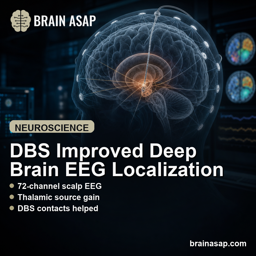
TL;DR: A 2026 study in Journal of Neural Engineering reported that adding passive recordings from bidirectional deep brain stimulation (DBS) electrodes to scalp electroencephalography (EEG) improved simulated deep-brain source localization, especially near the implanted lead.
Key Findings
- 72-channel scalp EEG: The simulations combined scalp EEG with 4-contact, 8-contact, or 40-contact DBS electrode configurations.
- Three noise levels: Source reconstruction was tested at 30 dB, 17.5 dB, and 5 dB signal-to-noise ratios.
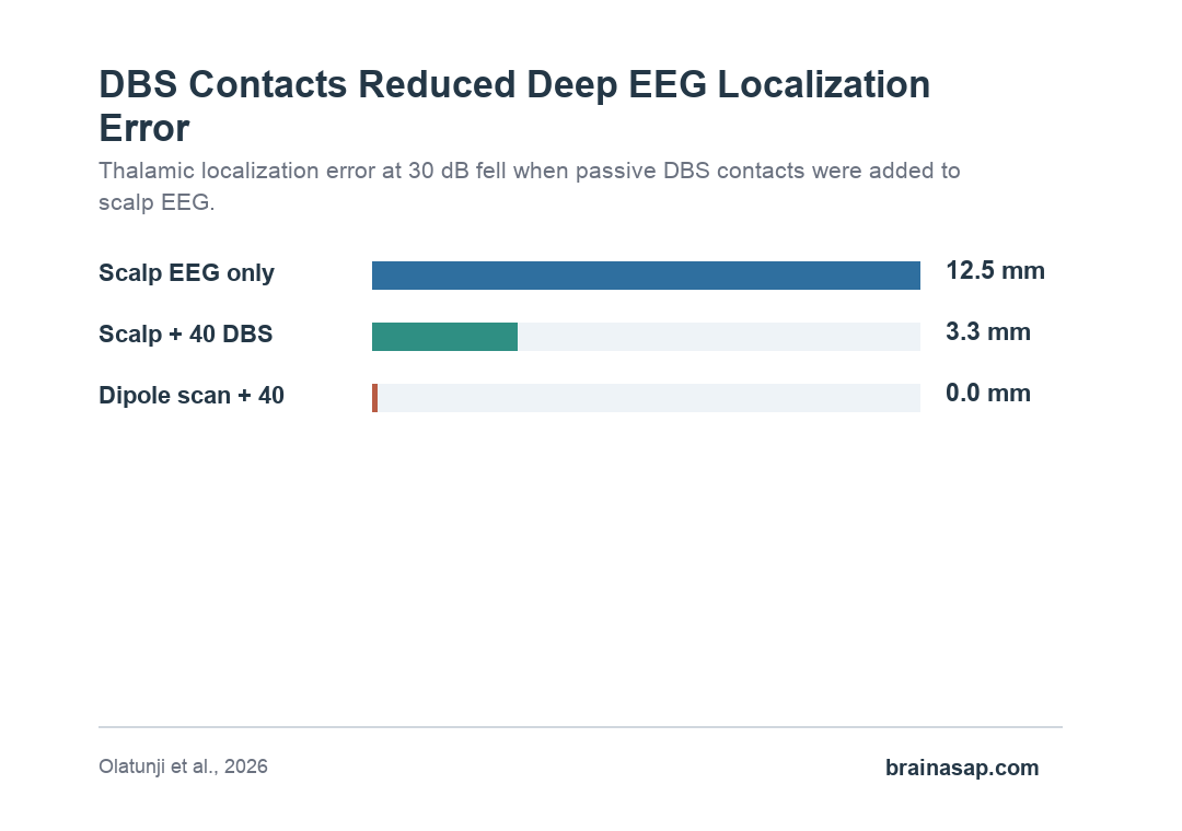
- Thalamic gain: At 30 dB, sLORETA thalamic error fell from 12.5 mm with scalp EEG alone to 3.3 mm with the 40-contact DBS setup.
- Dipole scan result: Dipole scan reached 0.0 mm average thalamic localization error at 30 dB with the 40-contact configuration.
- 8-contact tradeoff: The authors described the 8-contact configuration as the best balance between accuracy and electrode complexity.
Source: Journal of Neural Engineering (2026) | Olatunji et al.
Electroencephalography (EEG) records electrical activity from the scalp, but the inverse problem makes it difficult to identify where deep neural activity began.
This study asked whether patients who already have sensing-capable bidirectional DBS electrodes might offer an extra recording channel for localizing deep activity. The simulations focused on passive recording, not on stimulation-based probing.
The setup is therefore closest to a post-implantation monitoring problem, not a replacement for presurgical invasive mapping.
DBS Contacts Were Modeled as Passive Deep-Brain Recording Electrodes
The researchers built a finite element model that represented both scalp electrodes and implanted DBS contacts. They used an extended Complete Electrode Model, which accounted for electrode-tissue impedance and locally refined meshes around the implanted lead.
The electrode configurations were intentionally different in density:
- 4-contact DBS: A lower-density lead configuration.
- 8-contact DBS: A directional depth-electrode architecture closer to contemporary clinical sensing designs.
- 40-contact DBS: A high-density array used to test how much localization could improve with many contacts.
Each DBS setup was combined with 72-channel scalp EEG. Source reconstruction used standardized low-resolution electromagnetic tomography (sLORETA) and dipole scan methods across whole-brain, thalamus-focused, and hippocampus-focused analyses.
The clinical motivation was epilepsy and neuromodulation monitoring. In drug-resistant epilepsy, scalp EEG may miss early deep activity, while DBS electrodes can record local field potentials from chronically implanted targets.
Thalamic Localization Improved Most Near the Implanted DBS Lead
The clearest gains appeared in deep structures close to the invasive electrode. Under high recording quality at 30 dB SNR, sLORETA thalamic localization error fell from 12.5 mm with scalp EEG alone to 3.3 mm with the 40-contact DBS configuration.
At moderate recording quality, the dose-response pattern was stronger. Thalamic error decreased from 32.5 mm with scalp EEG alone to 12.8 mm with 4 contacts, 5.6 mm with 8 contacts, and 3.8 mm with 40 contacts.
At low recording quality, the same pattern remained but was less perfect:
- Scalp EEG alone: sLORETA thalamic error averaged 41.4 mm at 5 dB.
- 40-contact DBS plus scalp EEG: Error averaged 29.1 mm, a 29.7% improvement.
- Cingulate cortex: Error fell from 38.0 mm to 23.0 mm, a 39.5% reduction.
These numbers show why the location of the implanted lead is important. DBS contacts improved the local deep-source problem more than they changed ordinary cortical localization.
Dipole Scan Reached Near-Perfect Accuracy for Focal Sources
Dipole scan performed especially well for focal sources near the DBS lead. At 30 dB SNR, the 40-contact configuration reached 0.0 mm average thalamic localization error, compared with 3.3 mm for sLORETA in the same configuration.
In hippocampus-focused analysis, dipole scan showed a similar high-density advantage. With the 40-contact setup, left hippocampus error reached 0.0 mm at 30 dB and 0.6 mm at 17.5 dB.
The same method also reached 0.0 mm for the left amygdala at 30 dB.
- Focal-source strength: Dipole scan benefited most when the simulated source fit a compact single-source assumption.
- Distributed-source strength: sLORETA was described as more stable for broader regional analyses and two-source scenarios.
- Two-source cost: With scalp plus 40 contacts, dipole thalamic error changed from 0.0 mm to 0.6 mm when a second source was introduced.
The clinical boundary is narrow. The simulated physics favored multimodal recordings, especially when the deep source was close to the implanted lead and the inverse method matched the source pattern.
8-Contact DBS May Be the Practical Accuracy-Complexity Compromise
The 40-contact configuration usually produced the largest numerical gains, but the authors highlighted the 8-contact design as the best balance between accuracy and electrode complexity.
The density analysis showed the tradeoff. At 30 dB using sLORETA, moving from 4 contacts to 8 contacts improved thalamic localization accuracy by 31.1%.
Moving from 8 contacts to 40 contacts added another 21.4%, which suggests diminishing returns beyond the 8-contact design.
- Clinical fit: Modern sensing DBS systems are already implanted for therapy in some patients.
- Monitoring fit: Combined scalp and DBS recordings could support post-implantation epilepsy monitoring.
- Translation limit: The study was computational, so patient recordings and workflow validation remain necessary.
The study supports a narrow, useful claim: passive DBS recordings can add spatial information that scalp EEG lacks for deep sources. The next question is how reliably that advantage survives real patient movement, device noise, seizure variability, and clinical electrode placement.
Citation: DOI: 10.1088/1741-2552/ae87d0. Olatunji et al. Enhanced source localization accuracy through bidirectional deep brain stimulation (DBS) electrodes: A comparative study with non-invasive EEG methods. Journal of Neural Engineering. 2026;in press.
Study Design: Computational finite element source-localization simulation comparing scalp EEG alone with scalp EEG plus passive DBS electrode recordings.
Sample/Model: 72-channel scalp EEG combined with 4-contact, 8-contact, and 40-contact DBS configurations across 30 dB, 17.5 dB, and 5 dB SNR conditions.
Key Statistic: At 30 dB, sLORETA thalamic localization error fell from 12.5 mm with scalp EEG alone to 3.3 mm with scalp plus 40-contact DBS.
Caveat: The work was simulation-based and tested passive DBS recording, not a clinical trial of seizure localization or active stimulation probing.