How the Brain Hijacks Balance Control in Parkinson’s
TL;DR: When older adults face large balance challenges, their brains shift from relying on quick brainstem reflexes to slower cortical circuits—a shift that happens even in Parkinson’s disease, revealing a mechanistic window into age-related balance loss.
Balance isn’t automatic. When you stumble forward or feel the ground shift, your nervous system launches a cascade of muscle contractions that happens faster than conscious thought. But as bodies age and disease creeps in, this reflex arc starts to fail. The question neuroscientists have puzzled over: does the brain compensate by recruiting higher control centers, and if so, what does that tell us about why some older adults fall and others stay steady?
Key Findings
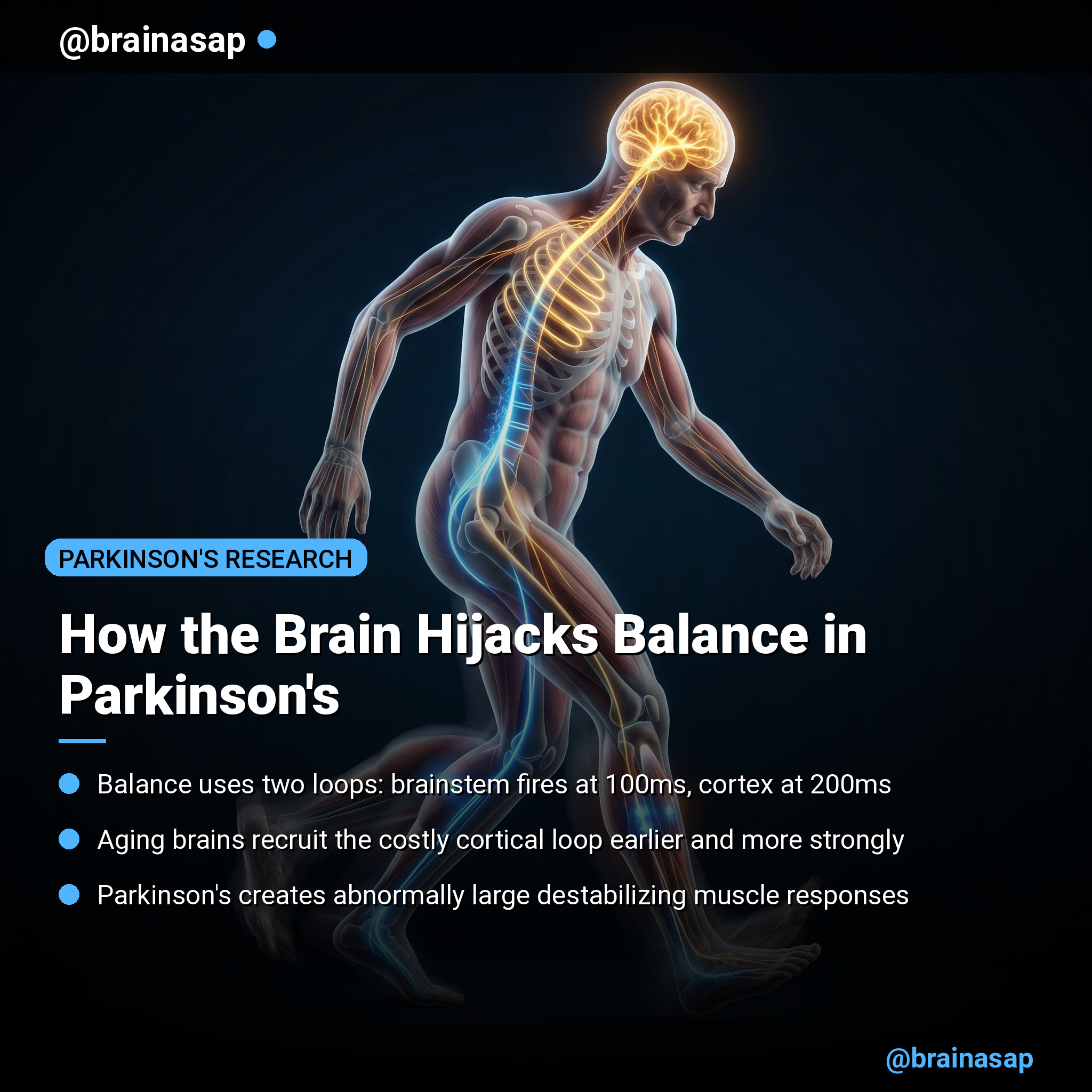
- Dual-pathway activation: Balance recovery uses two hierarchical feedback loops operating at different speeds—a brainstem loop at ~100-120ms and a cortical loop at ~200-210ms—allowing the nervous system to respond at multiple timescales.
- Challenge-driven cortical recruitment: As perturbations increase in magnitude, the longer-latency, cortically-mediated response grows stronger, but brainstem responses remain constant, showing how the brain recruits higher centers under pressure.
- Aging shifts the balance control hierarchy: Older adults show significantly larger cortical responses at lower perturbation magnitudes than younger adults, indicating the aging brain compensates with more cortical engagement earlier.
- Parkinson’s alters antagonist muscle control: People with Parkinson’s exhibit larger acceleration-driven destabilizing responses in opposing muscles, particularly in forward perturbations, suggesting basal ganglia dysfunction rewires how the brain dampens unwanted movement.
- Clinical correlations point to individual differences: In healthy older adults, worse balance ability correlates with abnormal destabilizing antagonist activity; in Parkinson’s, the same relationship breaks down, indicating disease transforms how muscle responses relate to fall risk.
- Double-loop model outperforms single-loop: Using a two-feedback-loop neuromechanical model improved reconstruction accuracy by ~10%, with R² values improving from 0.64-0.61 to 0.74-0.71 in healthy and Parkinson’s groups respectively.
Source: eNeuro (2026) | Boebinger et al.
The Two-Speed Reflex System That Keeps You Upright
For decades, neuroscientists viewed balance control as a straightforward brainstem operation. A disturbance hits your legs, stretch receptors fire, and the spinal cord triggers a corrective muscle contraction within 50 milliseconds—no brain involvement needed. But that model was incomplete.
Research over the past 15 years has shown that balance actually involves parallel feedback loops with different latencies and different jobs. The short-latency reflex (SLR) fires ~50ms after a perturbation—pure brainstem work, fast and dumb. But the long-latency reflex (LLR), arriving around 100-210ms later, requires cortical processing and carries information about the task goal, not just the sensory error.
The puzzle was always how to separate these components without brain recordings. Electromyography (EMG)—measuring muscle electrical activity—gives you one integrated signal that mixes both loops. This new study solved that by building a mathematical model that could decompose the EMG waveform into brainstem and cortical components based purely on their latencies.
Latencies Reveal the Neural Architecture
The researchers measured muscle responses in healthy older adults and people with Parkinson’s disease as they stood on a moving platform that jerked forward or backward at three magnitudes. They recorded the movements of their body and the electrical activity in the tibialis anterior (the shin muscle that resists forward falls) and the medial gastrocnemius (the calf, which resists backward falls).
Using center-of-mass kinematics and the muscle responses, they fitted a dual-loop neuromechanical model. The LLR1 component—consistent with brainstem mediation—appeared at approximately 100-120ms. The LLR2 component, lagging at 200-210ms, matched the latency expected from cortical processing and transmission.
The key insight: both loops existed in every participant. But their relative contribution shifted dramatically with challenge magnitude. Small perturbations? The brainstem loop handled it alone. Large perturbations? The cortex kicked in, amplifying the muscle response.
Aging Recruits Cortical Control Prematurely
The comparison between younger adults (published in prior work) and older adults revealed an age effect. Older adults engaged their cortical response (LLR2) at smaller perturbation magnitudes than younger adults—meaning they brought in the slower, more metabolically expensive cortical circuit earlier in the challenge spectrum.
Older adults with lower balance test scores tended to show larger destabilizing responses from their antagonist muscles—the muscles that should be quiet but, due to abnormal basal ganglia signaling, were firing inappropriately. In healthy aging, worse balance correlated with higher abnormal antagonist activity, suggesting a clear biomechanical liability.
What’s remarkable is that the cortical LLR2 component was similar between older adults and those with Parkinson’s disease. Contrary to the hypothesis that Parkinson’s would suppress cortical engagement, the disease didn’t dramatically change the LLR2 latency or the pattern of cortical recruitment with increasing challenge.
Parkinson’s Rewires the Antagonist Paradox
The antagonist muscle responses—the ones that should stabilize by counteracting the main corrective muscle—told a different story. In Parkinson’s disease, the destabilizing component of antagonist activity (driven by acceleration feedback) was abnormally large, especially in forward perturbations.
This aligns with the known motor phenotype of Parkinson’s: difficulty controlling movement scaling, excess muscle co-contraction, and abnormal force feedback. The basal ganglia, damaged by dopamine loss, usually fine-tune how much antagonist activity to engage. Without proper dopaminergic signaling, the antagonist muscle fires too aggressively, creating instability.
Curiously, this abnormal antagonist destabilizing activity did not correlate with clinical balance scores in the Parkinson’s group as strongly as in healthy older adults. This suggests that as Parkinson’s progresses, the neural-to-behavioral relationship becomes more complex—balance impairment may depend on multiple interacting deficits beyond just muscle response patterns.
A Window Into Balance Loss Without Brain Scans
One of the study’s most practical contributions is methodological. Researchers have traditionally inferred cortical engagement during balance by using fMRI or EEG alongside behavioral tasks. But those approaches are slow, noisy, and limited to lab settings.
This neuromechanical modeling approach extracts evidence of hierarchical neural control from muscle activity alone. By fitting the dual-loop model, researchers can estimate how much the cortex is contributing to balance recovery without ever recording cortical activity directly. The latency differences do the accounting.
This opens the door to assessing cortical involvement in balance clinically. If a patient’s balance responses show a shift toward larger cortical components at smaller challenges—or abnormal antagonist components—that could be diagnostic. Rehabilitation could be tailored: strengthen the circuits that are over-engaged, or address basal ganglia dopamine to fix the antagonist problem.
[Insert image: schematic showing dual-loop balance control—brainstem ~100ms and cortical ~200ms latencies with increasing perturbation magnitude]
Implications for Fall Prevention and Disease Tracking
Falls in older adults and people with Parkinson’s disease cost the healthcare system billions and devastate quality of life. Current assessments—balance tests, gait analysis—are behavioral readouts. They don’t tell you which neural systems are failing.
This study suggests that measuring muscle response patterns during reactive balance challenges could reveal the mechanistic culprits. In aging, cortical over-reliance on small challenges is a red flag. In Parkinson’s, abnormal antagonist destabilization is a specific biomarker. Both could guide interventions: cortical engagement might improve with targeted proprioceptive training; antagonist destabilization might respond to dopaminergic medications or deep brain stimulation.
The work also highlights why not all older adults fall despite similar age. Individual differences in how the hierarchy of balance control is engaged—whether the cortex kicks in early or the basal ganglia work smoothly—may predict who maintains balance and who doesn’t. Future studies could use these markers prospectively to identify high-risk individuals before a fall occurs.
Citation: Boebinger SC, Payne AM, Xiao J, Martino G, Borich MR, McKay JL, Ting LH. Cortically-mediated muscle responses to balance perturbations increase with perturbation magnitude in older adults with and without Parkinson’s disease. eNeuro. 2026;13(2):ENEURO.0423-25.2026. DOI: 10.1523/ENEURO.0423-25.2026
Authors’ affiliations: Wallace H. Coulter Department of Biomedical Engineering, Georgia Institute of Technology and Emory University, Atlanta, GA; Department of Physical Therapy, College of Health Sciences and Professions, Ohio University, Athens, OH; Department of Biomedical Sciences, University of Padova, Padua, Italy; Division of Physical Therapy and Department of Rehabilitation Medicine, Emory University, Atlanta, GA; Department of Neurology, Emory University, Atlanta, GA; Department of Biomedical Informatics, Emory University, Atlanta, GA.


