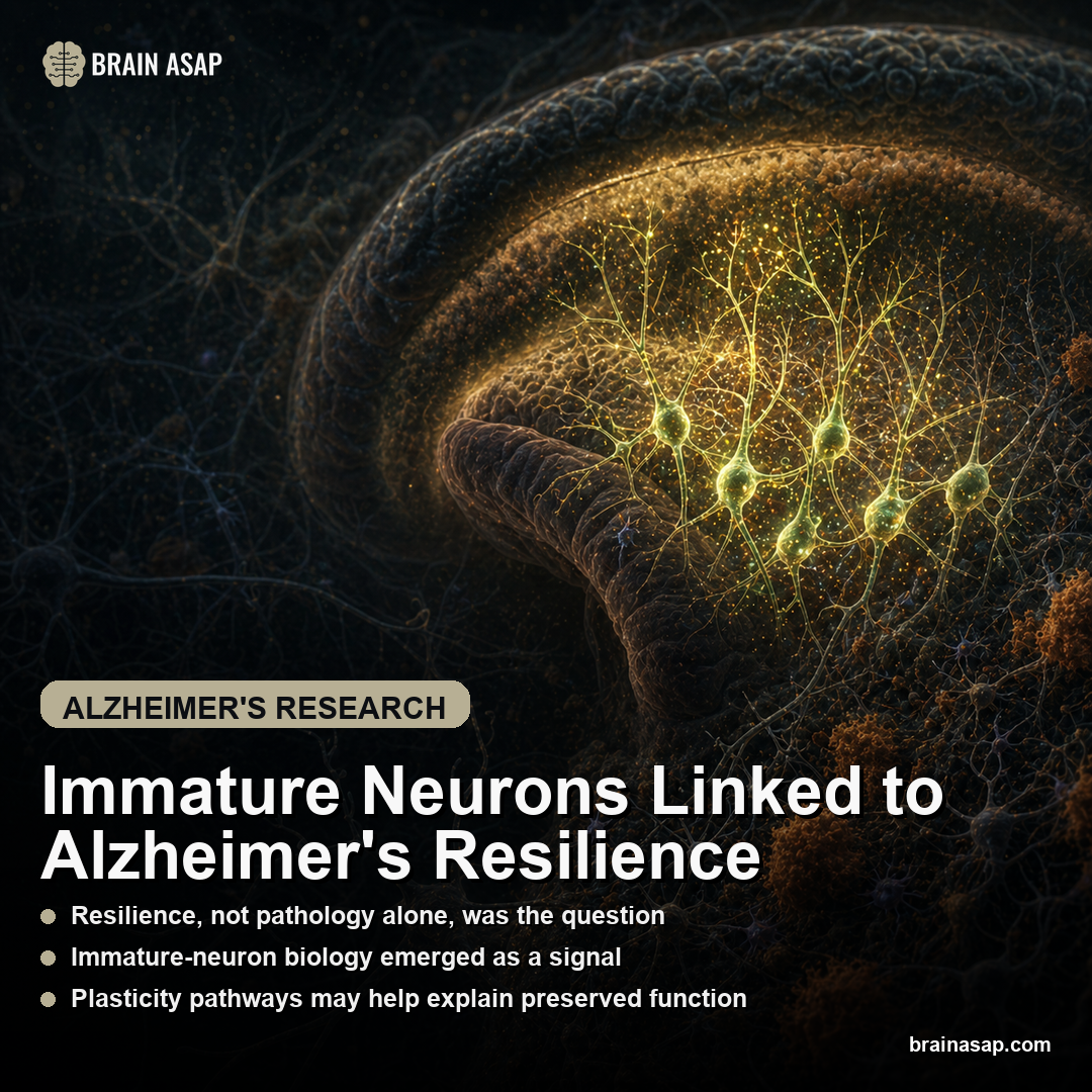
Immature Neurons Linked to Alzheimer’s Resilience
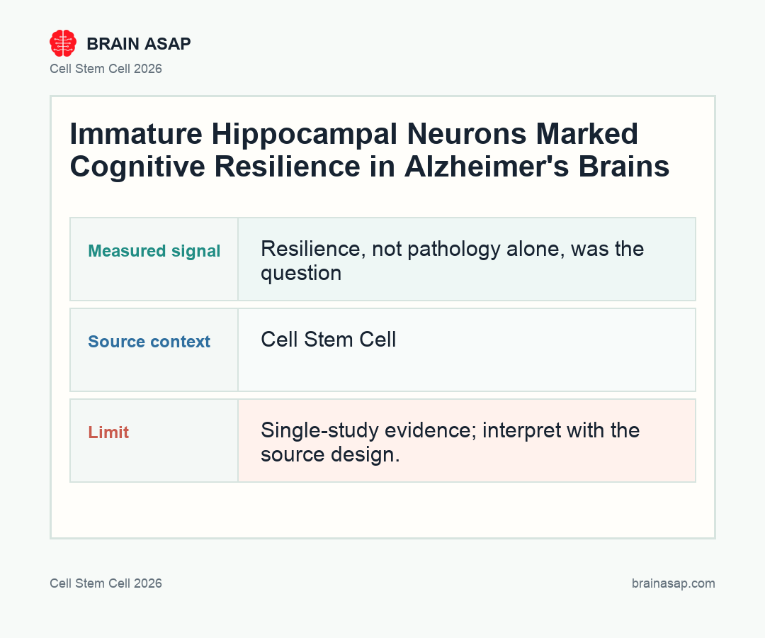
TL;DR: A 2026 Cell Stem Cell study used single-nucleus RNA sequencing of aged human hippocampus tissue and found rare immature neurons in healthy, Alzheimer’s, and dementia-resilient brains. The important signal was not simply whether these cells existed. It was how their gene activity changed in Alzheimer’s disease and in people who carried Alzheimer’s pathology but stayed cognitively resilient.
Key Findings
- The study compared three brain states: aged control donors, Alzheimer’s disease donors, and donors with Alzheimer’s pathology who remained dementia-resilient.
- The team focused on the hippocampus: this memory-related brain region is central to Alzheimer’s symptoms and is one of the main places scientists look for adult-born or immature neurons.
- Single-nucleus RNA sequencing was the main tool: this method reads gene activity from individual cell nuclei, allowing researchers to separate rare cell types that would be blurred together in bulk tissue.
- Immature neurons appeared across donor groups: the finding argues that these young-like neuronal populations can persist even in very old human brains.
- Resilience looked more like a cell-behavior question: the paper points toward differences in survival, inflammation, and “juvenile” cellular programs, not a simple count of how many immature neurons were present.
Source: Cell Stem Cell (2026) | Tosoni et al.
Alzheimer’s disease is usually described through damage: amyloid plaques, tau tangles, synapse loss, inflammation, and shrinking memory circuits. That damage is real, but it does not explain every person equally well.
Some older adults have substantial Alzheimer’s pathology in the brain and still avoid the level of cognitive impairment that would normally be expected.
This is called cognitive resilience. It means the brain is tolerating disease-related pathology better than expected. The new paper asks whether rare, young-looking neurons in the aged human hippocampus is plausibly part of that resilience system.
Those cells are called immature neurons because they show gene-expression patterns closer to developing neurons than fully mature adult neurons. That does not automatically mean the brain is replacing lost cells like a simple spare-parts machine. A more careful interpretation is that these cells can help preserve the local environment around memory circuits, especially when Alzheimer’s pathology is present.
Three Donor Groups Made Resilience the Main Comparison
The design was not just a healthy-versus-Alzheimer’s comparison. The researchers examined aged human hippocampus samples from three relevant groups:
- Aged control donors without Alzheimer’s pathology.
- Alzheimer’s disease donors with pathology and dementia.
- Dementia-resilient donors who had Alzheimer’s pathology but did not show the expected dementia syndrome.
That third group is the reason the paper is interesting. If a person has amyloid and tau pathology but remains cognitively intact, then pathology burden alone cannot be the whole explanation. The comparison lets researchers ask what the resilient brain is doing differently in the same disease environment.
The clean way to think about it is this: Alzheimer’s pathology is the pressure on the system. Cognitive resilience is the ability to keep functioning despite that pressure. This paper looks for cell-level signals that can help explain the difference.
Single-Nucleus RNA Sequencing Let the Team Find Rare Cells
The main technique was single-nucleus RNA sequencing, often shortened to snRNA-seq. RNA is the readout of which genes a cell is actively using. In this version of the method, researchers isolate nuclei from frozen tissue and measure gene activity one nucleus at a time.
The method is useful in postmortem human brain tissue because many important cell populations are rare. If researchers grind up a whole region and measure average gene activity, small populations can disappear into the average. With single-nucleus sequencing, the team can sort nuclei into cell types and cell states, including unusual neuron populations that look developmentally young.
The paper reports an integrated experimental and computational pipeline. In practical terms, the researchers did not rely on one marker and declare victory. They combined tissue sampling, sequencing, and data analysis to identify immature-neuron signatures in aged human hippocampus tissue.
This is especially important because adult human neurogenesis, the formation of new neurons in adulthood, has been debated for years. Rodent studies have strong evidence for adult-born hippocampal neurons, but human data have been harder to interpret. Human tissue quality, donor age, disease status, and marker choice can all change the result.
Immature Neurons Were Present Even in Very Old Hippocampus Tissue
The hippocampus is a memory-related structure deep in the temporal lobe. It is also one of the first brain regions people think about in Alzheimer’s disease because hippocampal damage is closely tied to memory decline.
In this study, immature neurons were detected across the donor groups. The finding is notable because these are not ordinary mature hippocampal neurons. They carry a more “juvenile” transcriptional profile, meaning their gene activity resembles younger or less fully developed neuronal states.
The phrase immature neuron can be misleading if it is read too loosely. It does not necessarily prove that large numbers of brand-new neurons are being born and wired into circuits.
It means the researchers found cells with immature neuronal signatures. Those signatures likely reflects adult-born neurons, delayed maturation, or a maintained young-like state in a small subset of cells.
That distinction is important because the therapeutic fantasy would be easy to oversell: make more young neurons, replace the damaged ones, and fix Alzheimer’s. The actual result is subtler. The cells may matter because of what they do locally, not because they replace every neuron that disease has harmed.
Cell Behavior Looked More Important Than Cell Count
The strongest interpretive point in the study is that resilience may depend less on whether immature neurons are present at all and more on how those cells behave under Alzheimer’s stress.
According to the research summary, the team expected that resilient brains might simply contain many more immature neurons than Alzheimer’s dementia brains. The difference was not that simple. Instead, the immature neurons in resilient tissue appeared to show gene programs linked with coping, survival, lower inflammation, and reduced cell-death signaling.
The result is biologically specific. A cell population can be present but ineffective, while another population can be small but active in a protective way.
In Alzheimer’s disease, where inflammation and network stress can spread through tissue, the state of a rare cell type may matter more than its raw number.
The authors connect these cells to homeostasis. Homeostasis means keeping the tissue environment stable enough for cells and circuits to function.
A resilient hippocampus does not necessarily be undamaged. It may be better at keeping the local environment from tipping into runaway dysfunction.
Juvenile Gene Programs can support the Surrounding Tissue
The paper describes the immature-neuron profiles as reflecting juvenile cellular functions that are compromised in Alzheimer’s disease. Here, “juvenile” means development-like gene activity: programs related to growth, flexibility, survival, and adaptation.
Memory circuits are living tissue, not just wires carrying information. Neurons need support from neighboring cells, blood vessels, immune cells, and molecular signals that help synapses stay functional.
If immature neurons release supportive signals or maintain a more flexible local state, they can help nearby circuits keep working longer.
This is not the same as saying immature neurons cure Alzheimer’s. The better interpretation is that these cells may be one piece of the resilience machinery. Other likely pieces include synapse maintenance, vascular health, immune regulation, education and cognitive reserve, sleep, metabolism, and the broader history of brain health across life.
Still, the cell-state result gives Alzheimer’s research a sharper target. Instead of only asking how to remove plaques or tangles, scientists can ask which cellular programs help an aged hippocampus tolerate pathology without collapsing into dementia.
The Treatment Angle Is Resilience, Not Simple Cell Replacement
The obvious treatment question is whether the protective cell programs can be strengthened. That is very different from injecting stem cells or trying to force the brain to make replacement neurons.
If the resilience signal is real, future therapies might aim to preserve the helpful gene programs inside immature neurons, reduce inflammatory signals that compromise them, or support the tissue conditions that let them function. Those are early research directions, not ready-made clinical instructions.
Alzheimer’s treatment is unlikely to be solved by one biological lever. Amyloid and tau remain central disease processes, but symptoms also depend on how the whole brain responds to pathology.
A therapy that reduces pathology and a therapy that improves resilience could eventually be complementary.
The Main Caveat Is That Postmortem Tissue Shows Associations
This human tissue study directly examined aged human hippocampus rather than relying only on mice. Postmortem tissue still has limits.
It can show which cell states are associated with Alzheimer’s disease and resilience. It cannot directly prove that immature neurons caused resilience in living people.
There are also measurement caveats. Cell-state labels depend on markers, tissue preservation, sampling location, sequencing depth, and computational decisions. Rare cells are especially difficult because small technical differences can influence what is detected.
The resilient Alzheimer’s brain appears to carry distinct immature-neuron transcriptional programs. These cells are important candidates for follow-up work, especially studies that test how they communicate with neighboring cells and whether their protective programs can be supported experimentally.
Alzheimer’s symptoms are not determined only by how much pathology is present. They also depend on whether the brain can preserve function in the presence of that pathology. This paper adds rare immature neurons in the aged hippocampus to the list of mechanisms that can help explain why some brains hold up better than others.
Paper: Transcriptional profiles of immature neurons in aged human hippocampus track Alzheimer’s pathology and cognitive resilience. Cell Stem Cell. 2026. DOI: 10.1016/j.stem.2026.04.002
Authors: Tosoni et al.
Design: Postmortem human hippocampus study using single-nucleus RNA sequencing and computational cell-state analysis.
Groups: Aged control donors, Alzheimer’s disease donors, and dementia-resilient donors with Alzheimer’s pathology.
Key Result: Immature-neuron populations persisted across donor groups, while their gene-expression programs tracked Alzheimer’s pathology and cognitive resilience.
Caveat: The study identifies associated cell states in human tissue. It does not prove by itself that immature neurons cause resilience or that boosting them would treat Alzheimer’s disease.